Abstract
Fetal cell microchimerism, the presence of genetically distinct fetal cells in maternal tissues, arises naturally during pregnancy when fetal stem cells migrate into maternal circulation. These cells can engraft in damaged tissues, including malignancies, and may play a role in tumor biology. This study evaluated the association between fetal microchimerism—detected through Testis-Specific Protein Y-encoded (TSPY) expression—and clinicopathologic features of invasive breast cancer in African American women. We conducted a blinded case–case study using immunohistochemical analysis of TSPY expression in 202 archival breast cancer tissues. TSPY protein, a marker of microchimeric Y-chromosome DNA, was assessed in relation to clinical and pathological characteristics including estrogen receptor (ER), progesterone receptor (PR), human epidermal growth factor receptor 2 (HER2) status, tumor grade, stage, size, and molecular subtype. Associations were examined using Chi-square tests. Survival outcomes were evaluated using Kaplan–Meier analysis. The results demonstrated that TSPY expression was not associated with patient age, receptor status, or triple-negative phenotype. However, TSPY positivity was significantly associated with higher tumor grade (p = 0.043), suggesting a relationship between fetal microchimerism and more aggressive disease features. Therefore, we concluded that microchimeric TSPY expression is enriched in higher-grade breast tumors among African American women. These findings suggest that fetal cell microchimerism may contribute to—or serve as a biomarker of—advanced breast cancer biology, although its mechanistic role requires further investigation.
|
Published in
|
Cancer Research Journal (Volume 14, Issue 3)
|
|
DOI
|
10.11648/j.crj.20261403.11
|
|
Page(s)
|
59-69 |
|
Creative Commons
|

This is an Open Access article, distributed under the terms of the Creative Commons Attribution 4.0 International License (http://creativecommons.org/licenses/by/4.0/), which permits unrestricted use, distribution and reproduction in any medium or format, provided the original work is properly cited.
|
|
Copyright
|
Copyright © The Author(s), 2026. Published by Science Publishing Group
|
Keywords
African American, Breast Cancer, Triple-negative, Microchimerism
1. Introduction
Fetal-derived stem cells have been identified in the breast stroma, where they can proliferate, differentiate, and integrate into maternal tissues
| [1] | Chan WF, Gurnot C, Montine TJ, Sonnen JA, Guthrie KA, Nelson JL, et al. Male microchimerism in the human female brain. PLoS ONE. 2012; 7(9): e45592. https://doi.org/10.1371/journal.pone.0045592 |
| [2] | Johnson K, Samura O, Nelson J, McDonnell W, Bianchi D. Significant fetal cell microchimerism in a nontransfused woman with hepatitis C: evidence of long-term survival and expansion. Hepatology. 2002; 36(5): 1295-1297.
https://doi.org/10.1053/jhep.2002.36798 |
| [3] | Yin Y, Li Y, Qiao H, Wang H, Yang X, Zhang H, Pang X, Zhang Y, Chen W. TSPY is a cancer testis antigen expressed in human hepatocellular carcinoma. British Journal of Cancer. 2005; 93(4): 458-463.
https://doi.org/10.1038/sj.bjc.6602710 |
| [4] | Zeng X, Tan K, Yeo A, Sasajala P, Tan X, Xiao ZC, et al. Pregnancy-associated progenitor cells differentiate and mature into neurons in the maternal brain. Stem Cells and Development. 2010; 19(12): 1819-1830.
https://doi.org/10.1089/scd.2010.0046 |
[1-4]
. This phenomenon, known as fetal cell microchimerism, occurs naturally during pregnancy and results in the long-term persistence of genetically distinct fetal cells within the maternal host
. These cells have been detected in both normal and malignant breast tissues, suggesting a potential role in tissue remodeling and tumor biology.
Microchimeric cells have been hypothesized to contribute to the extensive epithelial expansion required for lactation during pregnancy, as well as to processes involved in tumor initiation and progression
| [6] | Geck P. Symptotic detection of chimerism: Y does it matter? Chimerism. 2013; 4(4): 144-146.
https://doi.org/10.4161/chim.26945 |
| [7] | Dhimolea E, Denes V, Lakk M, Al-Bazzaz S, Aziz-Zaman S, Pilichowska M, Geck P. High male chimerism in the female breast shows quantitative links with cancer. International Journal of Cancer. 2013; 133(4): 835-842.
https://doi.org/10.1002/ijc.28065 |
[6, 7]
. Consequently, microchimerism may provide a biologically plausible mechanism underlying tumor aggressiveness, therapeutic resistance, and the high prevalence of triple-negative breast cancer (TNBC) among African American women.
Breast cancer remains a major public health concern, accounting for over 40,000 deaths annually in the United States
. Importantly, its clinical behavior varies across racial and ethnic groups. African American women are disproportionately affected by more aggressive disease, often presenting with higher-grade tumors, advanced-stage disease, and subtypes lacking targetable receptors
. Breast cancer is a heterogeneous disease composed of multiple intrinsic molecular subtypes identified through gene expression profiling
| [11] | Perou C, Jeffrey S, van de Rijn M, Rees C, Eisen M, Ross D, et al. Distinctive gene expression patterns in human mammary epithelial cells and breast cancers. Proceedings of the National Academy of Sciences of the United States of America. 1999; 96: 9212-9217. https://doi.org/10.1073/pnas.96.16.9212 |
| [12] | Sorlie T, Perou C, Tibshirani R, Aas T, Geisler S, Johnsen H, et al. Gene expression patterns of breast carcinomas distinguish tumor subclasses with clinical implications. Proceedings of the National Academy of Sciences of the United States of America. 2001; 98: 10869-10874.
https://doi.org/10.1073/pnas.191367098 |
| [13] | Hall M, Reid J, Burbidge L, Pruss D, Deffenbaugh A, Frye C, et al. BRCA1 and BRCA2 mutations in women of different ethnicities undergoing testing for hereditary breast-ovarian cancer. Cancer. 2009; 115: 2222-2233.
https://doi.org/10.1002/cncr.24200 |
[11-13]
. Among these, TNBC—defined by the absence of estrogen receptor (ER), progesterone receptor (PR), and HER2 expression is particularly aggressive and associated with poor prognosis due to limited therapeutic options
| [14] | Carey L, Winer E, Viale G, Cameron D, Gianni L. Triple-negative breast cancer: disease entity or title of convenience? Nature Reviews Clinical Oncology. 2010; 7: 683-692.
https://doi.org/10.1038/nrclinonc.2010.154 |
| [15] | Perou CM, Sørlie T, Eisen MB, van de Rijn M, Jeffrey SS, Rees CA, et al. Molecular portraits of human breast tumours. Nature. 2000; 406(6797): 747-752. https://doi.org/10.1038/35021093 |
[14, 15]
.
The role of microchimerism in breast cancer remains controversial. Some studies suggest that these cells may exert protective or reparative effects by contributing to tissue regeneration
, while others propose a tumorigenic role, potentially influencing cancer initiation and progression
| [18] | Kalluri R, Zeisberg M. Fibroblasts in cancer. Nature Reviews Cancer. 2006; 6(5): 392-401. https://doi.org/10.1038/nrc1877 |
| [19] | Lee E, Seppanen SM, Bou-Gharios G, Seppanen E, Khosrotehrani E, Fisk N. Fetal stem cell microchimerism: natural-born healers or killers? Molecular Human Reproduction. 2010; 16(11): 869-878. https://doi.org/10.1093/molehr/gaq060 |
[18, 19]
. Clarifying this duality requires evaluation of microchimerism in relation to tumor subtype and established prognostic factors.
In this study, we evaluated fetal microchimerism in breast tumors from African American women using immunohistochemical detection of testis-specific protein Y-encoded (TSPY), a marker of Y-chromosome-derived cells. We demonstrate that TSPY expression is significantly associated with higher tumor grade and shows an increasing trend with tumor size, although no association was observed with receptor status. These findings suggest that microchimerism may serve as a biomarker of aggressive breast cancer in this population.
2. Materials and Methods
2.1. Study Design
This study was reviewed and deemed exempt by the Howard University Institutional Review Board (IRB-15-MED-19). A total of 202 cases of invasive ductal carcinoma (IDC) from African American women diagnosed and treated at Howard University Hospital between 2000 and 2010 were included. Cases were selected based on the availability of adequate tumor tissue. Clinical and demographic data were obtained from the Howard University Cancer Center Tumor Registry.
2.2. Tissue Samples and Microarray Construction
Formalin-fixed, paraffin-embedded (FFPE) tumor specimens were used to construct tissue microarrays (TMAs) (Pantomics, Inc., Richmond, CA). Each TMA consisted of 1.0 mm cores sampled from representative tumor regions, with two cores per case. Sections (5 µm) were cut and mounted on microscope slides. Normal FFPE prostate tissue served as a positive control for TSPY expression.
2.3. Detection of Microchimerism
Microchimerism was assessed using immunohistochemistry (IHC) targeting TSPY, a Y-chromosome-encoded protein present in male cells. TSPY is highly repetitive within the Y chromosome and is considered a sensitive and reliable marker for detecting male microchimeric cells in female tissues
| [7] | Dhimolea E, Denes V, Lakk M, Al-Bazzaz S, Aziz-Zaman S, Pilichowska M, Geck P. High male chimerism in the female breast shows quantitative links with cancer. International Journal of Cancer. 2013; 133(4): 835-842.
https://doi.org/10.1002/ijc.28065 |
| [20] | Ali S. Genetic integrity of the human Y-chromosome exposed to groundwater arsenic. BMC Medical Genomics. 2010; 3(1): 35. https://doi.org/10.1186/1755-8794-3-35 |
| [21] | Li Y, Tabatabi Z, Lee T, Hatakeyama S, Ohyama C, Chan W, Looijenga L, et al. The Y-encoded TSPY protein: a significant marker potentially plays a role in the pathogenesis of testicular germ cell tumors. Human Pathology. 2007; 38(10): 1470-1481. https://doi.org/10.1016/j.humpath.2007.02.014 |
[7, 20, 21]
.
2.4. Immunohistochemistry
Slides were deparaffinized, rehydrated, and subjected to heat-induced antigen retrieval (pH 9.0). Staining was performed using a rabbit polyclonal anti-TSPY antibody (Lifespan Biosciences) at an optimized concentration of 0.01 µg/mL on a Dako Autostainer system.
2.5. Scoring
TSPY expression was evaluated independently by two pathologists blinded to clinical data. An H-score system was used, with scores ≥200 classified as positive and <200 as negative.
2.6. Breast Cancer Subtypes
Tumors were classified into molecular subtypes based on ER, PR, HER2, and Ki-67 expression, following established criteria
| [12] | Sorlie T, Perou C, Tibshirani R, Aas T, Geisler S, Johnsen H, et al. Gene expression patterns of breast carcinomas distinguish tumor subclasses with clinical implications. Proceedings of the National Academy of Sciences of the United States of America. 2001; 98: 10869-10874.
https://doi.org/10.1073/pnas.191367098 |
| [22] | Paik S. Molecular profiling of breast cancer. Current Opinion in Obstetrics and Gynecology. 2006; 18: 59-63.
https://doi.org/10.1097/01.gco.0000192983.19143.2e |
[12, 22]
.
2.7. Reagents
Antibodies used in this study were as follows: TSPY, and β-tubulin (Santa Cruz Biotechnology, Dallas, TX).
2.8. Culture of Breast and Prostate Cancer Epithelial Cell Lines
HCC70 (African American breast cancer), HCC1806 (African American breast cancer), and 22RV1 (metastatic resistant prostate cancer) epithelial cells were obtained from American Type Culture Collection (ATCC) (Manassas, VA). All three cell lines were maintained using advanced RPMI-1640 supplemented with 10% Fetal Bovine Serum (FBS), 100 U/ml penicillin, and 100mg/ml streptomycin at 37°C in a 5% CO2 atmosphere.
2.9. Western and Protein Isolation
HCC70, HCC1806, and 22RV1 epithelial cancer cells were cultured at 800,00 cells in 60 mm dishes for 24 hr. After 24 hr. protein cell lysates were collected with Solulyse-M Protein Extraction Reagent [a non-ionic detergent, 250 mM sucrose and 25mM Tris; pH 7.4] (Gentalis, San Diego, CA). Complete Mini Protease Inhibitor Cocktail was added to the lysis buffer. Protein extraction was performed according to the manufacturer’s protocol. A total of 30 ug of protein was used for SDS-PAGE. 4-20% polyacrylamide gels (Biorad, Hercules, CA) were used to resolve proteins in SDS tris-glycine running buffer (1X) containing 25mM Tris, 192 mM glycine, and 0.1% (w/v) SDS, pH 8.3 (Biorad, Hercules, CA). The voltage was set to 100-115 volts to resolve the proteins. The proteins were transferred using an iBlot Transfer Device (LifeTechnologies, Carslbad, CA), which performed a dry transfer in 7 minutes at 20 volts. A polyvinylidene difluoride (PVDF) membrane located in iBlot Transfer Stacks (Life Technologies, Carslbad, CA) was used. Blocking buffer, antibody wash solution, primary diluent, and secondary diluent were prepared from components of WesternBreeze Chemiluminescent Immunodetection Kit (LifeTechnologies, Carslbad, CA) according to the manufacture’s protocol. After the transfer was complete, membranes were blocked for 1 hour in 10 mL of blocking buffer at room temperature. Incubation of the membrane in blocking buffer and all other solutions were performed on a rocker at 60 rpm. Prior to the addition of diluted primary antibody, the membranes were washed 2 times for 2 minutes with distilled water. Membranes were incubated with primary antibody for an hour at room temperature or overnight at 4ºC. Membranes were washed 4 times for 5 minutes after primary antibody incubation period in antibody wash, followed by incubation with 10 mL of secondary antibody conjugated to horseradish peroxidase (HRP) (1:10,000) or alkaline phosphatase for 1 hour at room temperature. Anti-goat, anti-rabbit or anti-mouse alkaline phosphatase conjugated secondary antibodies were pre-made and provided in WesternBreeze Chemiluminescent Immunodection Kit. After secondary antibody incubation period, the membranes were washed with antibody wash 4 times for 5 minutes and then 2 times with distilled water before adding the appropriate chemiluminescent substrate. Supersignal West Femto Chemiluminescent substrate (Pierce of Thermo Scientific, Rockford, IL) was used for detection of secondary antibody conjugated to HRP. CDP-Star, from WesternBreeze Chemiluminescent Immunodetection Kit (Lifetechnolgies, Carlsbd, CA), was used as a substrate for secondary antibodies conjugated to alkaline phosphatase. Protein band detection and densitometry was acquired with ChemiDoc XRS+ Image Lab software (Biorad, Hercules, CA) using a charged coupled device (CCD) camera imager.
3. Statistical Analysis
Associations between TSPY expression and clinicopathologic variables were assessed using Chi-square tests. Survival analyses were conducted using Kaplan–Meier curves and log-rank tests. Statistical analyses were performed using SPSS version 22, with significance set at p < 0.05.
4. Results
4.1. TSPY Protein Expression Across Breast Cancer Histologies
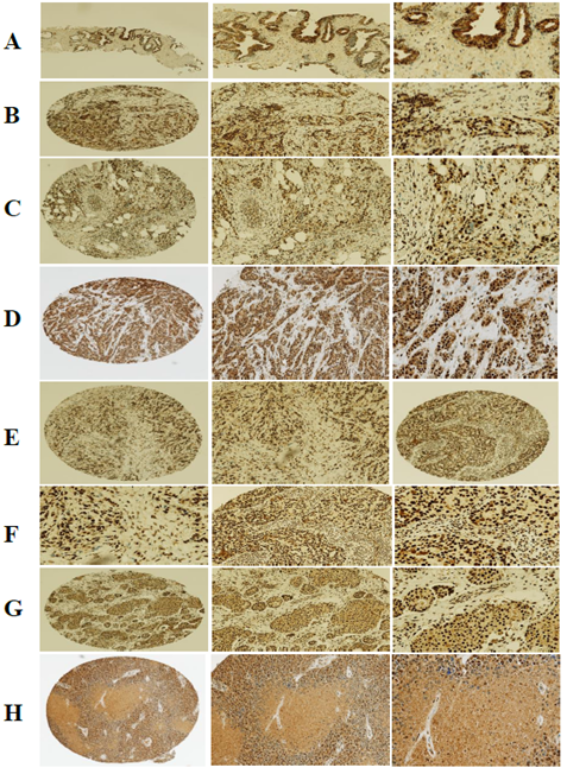
TSPY protein expression was evaluated in control and tumor tissues, as illustrated in
Figure 1. Positive staining was confirmed in normal prostate gland tissue (
Figure 1A), validating assay specificity.
Across breast cancer specimens, TSPY expressions were observed in multiple histologic subtypes and tumor grades. High levels of TSPY expression were identified in grade 2 luminal A invasive ductal carcinoma (IDC) with calcifications and granulomatous features (
Figure 1C), as well as in grade 2 luminal A (IDC) (
Figure 1D). Elevated expression was also evident in more aggressive disease, including grade 3 IDC with Paget’s disease (
Figure 1E), grade 3 IDC with dense stromal fibrosis (
Figure 1F), and grade 3 triple-negative IDC (
Figure 1G).
Additionally, TSPY positivity was observed in grade 1 DCIS/IDC with fibrocystic changes and apocrine metaplasia, indicating that microchimerism may be present across both early and advanced lesions. In contrast, grade 3 triple-negative IDC with sclerosing adenosis demonstrated low or absent TSPY expression (
Figure 1H), highlighting heterogeneity in microchimeric cell distribution.
Figure 1. TSPY protein expression in normal prostate and breast cancer tissues of varying grades and molecular subtypes. A) Normal Prostate Gland; B-C) Grade 2 Luminal A IDC; D) Grade 3 IDC with Pagets disease;. E) Grade 3 IDC Dense stromal fibrosis; F) Grade 3 triple negative IDC; G) Grade 1 DCIS/IDC; H) Grade 3 triple negative IDC with sclerosing adenosis. Representative images are shown at 4×, 10×, and 20× magnification.
4.2. Association Between TSPY Expression and Clinicopathologic Features
Associations between TSPY expression and clinicopathologic parameters are summarized in
Table 2. A statistically significant association was observed between TSPY expression and higher tumor grade (p = 0.043). Mean microchimeric cell counts increased from 243.17 ± 47.06 cells in grade 1–2 tumors to 257.60 ± 31.91 cells in grade 3 tumors, supporting a link between microchimerism and tumor aggressiveness.
Table 1. Clinical and pathologic characteristics of study population with Breast Cancer.
Population | Description | N | % |
Age (years) | | | |
| <50 | 57 | 28.2 |
| >50 | 145 | 71.8 |
ER status | | |
| Positive | 49 | 39.2 |
| Negative | 76 | 60.8 |
PR status | | |
| Positive | 60 | 48.0 |
| Negative | 65 | 52.0 |
HER2 status | | | |
| Positive | 112 | 89.6 |
| Negative | 13 | 10.4 |
Subtype* | | | |
| Luminal A | 62 | 49.6 |
| Luminal B | 14 | 11.2 |
| Her2 | 8 | 6.4 |
| Triple-negative | 41 | 32.8 |
Stage at diagnosis | | |
| I | 37 | 30.1 |
| II | 50 | 40.7 |
| III | 27 | 22.0 |
| IV | 9 | 7.3 |
Size | <20mm | 47 | 37.6 |
| 20-50mm | 42 | 33.6 |
| >50mm | 19 | 15.2 |
| Unknown | 17 | 13.6 |
Grade | | | |
| 1 and 2 | 41 | 32.8 |
| 3 | 84 | 67.2 |
Distant Mets | | | |
| None | 105 | 84.0 |
| Distant Mets | 20 | 16.0 |
Lymph Node Status | | | |
| Negative | 59 | 57.3 |
| Positive | 44 | 42.7 |
Luminal | | | |
| Not luminal | 49 | 39.2 |
| Luminal | 76 | 60.8 |
Triple Negative | | | |
| Not triple negative | 83 | 66.4 |
| Triple negative | 42 | 33.6 |
Table 2. Association of TSPY expression with clinicopathologic parameters of Breast Cancer.
Breast Cancer Parameter | Description | TSPY Negative | TSPY Positive | P-value* |
ER Status | | | | |
| Negative | 6 | 43 | |
| Positive | 8 | 68 | .491 |
PR Status | | | | |
| Negative | 7 | 53 | |
| Positive | 7 | 58 | .548 |
HER2 | | | | |
| Negative | 14 | 98 | |
| Positive | 0 | 13 | .196 |
Triple Negative | | | | |
| No | 8 | 75 | |
| Yes | 6 | 36 | .310 |
Distant Metastases | | | | |
| No | 12 | 93 | |
| Yes | 2 | 18 | .506 |
Subtype | | | | |
| Luminal A | 8 | 54 | |
| Luminal B | 0 | 14 | |
| Her2+ | 0 | 8 | |
| Triple-negative | 6 | 35 | .328 |
Stage at Diagnosis | | | | |
| I | 4 | 33 | |
| II | 7 | 43 | |
| III | 3 | 24 | |
| IV | 0 | 9 | 0.680 |
Size | | | | |
| <20mm | 4 | 43 | |
| 20-50mm | 6 | 36 | |
| >50mm | 0 | 19 | .199 |
Grade | | | | |
| 1+2 (ref) | 8 | 33 | |
| 3 | 6 | 78 | .043 |
Lymph Nodes | | | | |
| Negative | 8 | 51 | |
| Positive | 4 | 40 | .353 |
Luminal | | | | |
| Not Luminal | 6 | 43 | |
| Luminal | 8 | 68 | .491 |
Tumor Size | | | | |
| T1 | 4 (40.0%) | 43 (43.9%) | |
| T2 | 6 (60.0%) | 36 (36.7%) | |
| T3 | 0 (0%) | 19 (19.4%) | 0.19 |
An increasing trend in microchimeric cell counts was also observed with tumor size, with mean values of 253.40 ± 44.42 cells for T1 tumors (<20 mm), 254.76 ± 36.59 cells for T2 tumors (20–50 mm), and 260.95 ± 21.94 cells for T3 tumors (>50 mm). However, this trend did not reach statistical significance (p = 0.199).
No significant associations were found between TSPY expression and ER status (p = 0.491), PR status (p = 0.548), HER2 status (p = 0.196), triple-negative status (p = 0.310), luminal subtype (p = 0.491), stage at diagnosis (p = 0.680), lymph node status (p = 0.353), or distant metastasis (p = 0.506). Mean microchimeric cell counts were 249.66 ± 45.08 cells in node-positive cases and 247.15 ± 27.63 cells in cases with distant metastasis.
4.3. Validation of the TSPY expression in breast cancer epithelial cells
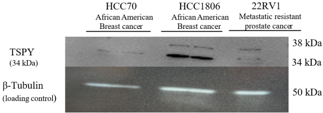
To validate TSPY protein expression, we performed Western blot analysis using two African American breast cancer epithelial cell lines, HCC70 and HCC1806, with the 22RV1 castration-resistant prostate cell line serving as a positive reference control for TSPY expression (
Figure 2). The HCC70 cell line was established from a 49-year-old Black female patient diagnosed with ductal carcinoma, whereas the HCC1806 cell line derived from a 60-year-old Black Female patient with primary acantholytic squamous cell carcinoma. The 22RV1 cell line, derived from a relapsed prostate tumor, is a well-established model of metastatic castration-resistant prostate cancer and was included as a comparator for TSPY expression.
Figure 2. Representative Western blot showing TSPY protein expression in HCC70, HCC1806, and 22RV1 cell lines. TSPY expression varied among cell lines, with highest expression in HCC1806, moderate expression in 22RV1, and minimal expression in HCC70. β-Tubulin served as the loading control.
Western blot analysis demonstrated differential TSPY expression among the three cell lines. HCC1806 exhibited robust TSPY protein expression, whereas HCC70 displayed minimal expression. Similarly, the 22RV1 control cell line showed relatively low levels of TSPY expression. Equal protein loading was confirmed by total protein staining and β-Tubulin. These findings validate the expression of TSPY in advanced breast cancer epithelial cells and identify HCC1806 as a TSPY-expressing African American breast cancer model, supporting its utility for future mechanistic studies investigating the role of TSPY in breast cancer progression and tumor biology.
4.4. Survival Analysis
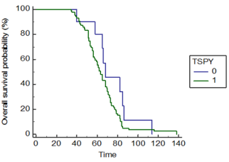
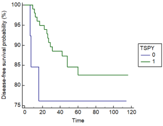
Overall survival (OS) and disease-free survival (DFS) were evaluated using Kaplan–Meier analysis (
Figures 3 and 4). Median OS was 68.0 months (96% CI: 65.0–86.0) for tumors with low TSPY expression and 63.0 months (95% CI: 58.0–68.0) for TSPY-positive tumors, with no statistically significant difference observed (p = 0.15) (
Figure 2).
Figure 3. Image of Kaplan-Meier analysis of overall survival by TSPY expression (negative = 0, positive = 1) in breast cancer.
Similarly, TSPY expression was not significantly associated with DFS (p = 0.21) (
Figure 3). The mean DFS was 89.17 months (95% CI: 64.57–113.77) in TSPY-negative tumors and 101.2 months (95% CI: 94.30–108.08) in TSPY-positive tumors.
Figure 4. Kaplan–Meier analysis of disease-free survival by TSPY protein expression in breast cancer (recurrence = 1, no recurrence = 0).
5. Discussion
This study demonstrates that TSPY-detected microchimerism is significantly associated with higher tumor grade in breast cancer among African American women, a population disproportionately affected by aggressive disease. While prior studies have linked microchimerism to tumor presence, our findings extend this body of work by identifying a quantitative and statistically significant relationship between TSPY expression and tumor grade, suggesting a role in tumor progression rather than passive tissue integration
| [7] | Dhimolea E, Denes V, Lakk M, Al-Bazzaz S, Aziz-Zaman S, Pilichowska M, Geck P. High male chimerism in the female breast shows quantitative links with cancer. International Journal of Cancer. 2013; 133(4): 835-842.
https://doi.org/10.1002/ijc.28065 |
| [23] | Dubernard G, Oster M, Chareyre F, Antoine M, Rouzier R, Uzan S, et al. Increased fetal cell microchimerism in high grade breast carcinomas occurring during pregnancy. International Journal of Cancer. 2009; 124(5): 1054-1059.
https://doi.org/10.1002/ijc.24081 |
[7, 23]
. These findings are consistent with emerging evidence that pregnancy-acquired microchimerism represents a biologically important component of the tumor microenvironment. A recent systemic review and meta-analysis demonstrated that male-origin microchimerism is significantly associated with cancer risk while emphasizing substantial heterogeneity across tumor types and the need for tissue-based mechanistic studies
. Likewise, contemporary reviews suggest that fetal microchimeric cells may exert both tumor-protective and tumor-promoting effects depending on immune context, tissue environment, and cellular differentiation state
| [25] | Parmeggiani C, Sallinger K, Cleaves HJ II, et al. The duality of microchimerism and cancer in parous women: a review and evolutionary perspective. Seminars in Immunopathology. 2025; 47: 15. https://doi.org/10.1007/s00281-025-01041-0 |
[25]
.
A key contribution of this study is the focus on testis-specific protein Y-encoded (TSPY) as both a biomarker and a potential functional mediator. Increasing evidence indicates that TSPY plays an active role in oncogenesis. TSPY has been shown to regulate cell cycle progression through interactions with cyclin B/CDK1, promote cellular proliferation, and influence transcriptional programs associated with tumor growth
. More recent studies further suggest that aberrant TSPY expression contributes to tumorigenesis, genomic instability, and oncogenic signaling pathways in cancers such as prostate, hepatocellular carcinoma, and germ cell tumors
. These findings support a model in which TSPY-positive microchimeric cells may actively participate in tumor-promoting processes within the breast tumor microenvironment. Although most studies of TSPY have focused on prostate, germ cell, and hepatocellular malignancies, evidence indicates that cancer-testis antigens contribute to tumor plasticity, immune escape, and genomic instability across multiple solid tumors
. Our findings suggest that TSPY may similarly identify biologically active microchimeric cells within aggressive breast cancers, extending its potential role beyond traditionally recognized TSPY-associated malignancies.
Importantly, TSPY expression in this cohort was not associated with ER, PR, HER2, or triple-negative status, suggesting that its relationship with tumor grade is independent of receptor-defined molecular subtypes. This observation implies that microchimerism may influence core tumorigenic processes, including proliferation, stromal remodeling, and immune modulation, rather than receptor-mediated signaling pathways. The observed trend toward increasing microchimeric cell counts with tumor size further supports a potential role in disease progression, although causality cannot be established from the current study.
The biological role of fetal microchimeric cells in cancer remains complex and is increasingly recognized as context dependent. While early investigations reported conflicting associations between fetal microchimerism and breast cancer, recent evidence suggests that these cells may function as immune modulators, tissue repair progenitors, or contributors to tumor progression depending on the local microenvironment. A recent meta-analysis concluded that male-origin microchimerism demonstrates a significant association with cancer overall, while emphasizing the need to characterize the phenotype and functional properties of tissue-resident microchimeric cells
. Similarly, Parmeggiani et al. proposed that fetal microchimeric cells may transition between protective and tumor-promoting phenotypes in response to inflammatory and neoplastic signals
| [25] | Parmeggiani C, Sallinger K, Cleaves HJ II, et al. The duality of microchimerism and cancer in parous women: a review and evolutionary perspective. Seminars in Immunopathology. 2025; 47: 15. https://doi.org/10.1007/s00281-025-01041-0 |
[25]
. Prior studies have demonstrated reduced circulating microchimerism in peripheral blood of breast cancer patients, suggesting recruitment and localization of these cells within tumor tissue
. One hypothesis is that tumors actively recruit microchimeric cells via chemokine-mediated homing mechanisms, similar to stem cell trafficking. Alternatively, microchimeric cells may initially contribute to tissue repair but undergo functional reprogramming within the tumor microenvironment, adopting pro-tumorigenic phenotypes that support angiogenesis, immune evasion, or stromal expansion
. In this context, TSPY expression may identify a subset of microchimeric cells with enhanced proliferative or oncogenic potential.
These findings are particularly relevant to breast cancer disparities. Understanding the contribution of pregnancy-associated microchimerism to aggressive breast cancer may provide new insight into biologic mechanisms underlying disparities that are not explained by conventional molecular subtypes alone. Recent reviews have emphasized the need to investigate microchimerism across diverse populations because most published studies have involved predominately European cohorts
| [25] | Parmeggiani C, Sallinger K, Cleaves HJ II, et al. The duality of microchimerism and cancer in parous women: a review and evolutionary perspective. Seminars in Immunopathology. 2025; 47: 15. https://doi.org/10.1007/s00281-025-01041-0 |
[25]
. African American women are more likely to develop high-grade, treatment-resistant tumors, yet the underlying biological drivers remain incompletely understood
. Although TSPY expression was not associated with triple-negative status, its correlation with tumor grade suggests that it may serve as a complementary biomarker of aggressive disease, particularly in cases where traditional receptor-based classification provides limited prognostic value. Integration of microchimerism markers such as TSPY into existing biomarker panels may improve risk stratification and guide therapeutic decision-making.
From a translational perspective, TSPY and microchimerism represent promising areas for further investigation. TSPY detection could be incorporated into multiplex diagnostic platforms alongside established biomarkers. Moreover, if functional studies confirm a causal role, TSPY-associated pathways may provide novel therapeutic targets, particularly for aggressive tumors lacking conventional receptor targets. Additionally, characterization of microchimeric cell populations may offer new insights into tumor–host interactions, an emerging area of importance in cancer biology and immuno-oncology.
Several limitations should be considered. The use of Y-chromosome-based detection restricts analysis to male-derived fetal cells, excluding microchimerism from female pregnancies. The absence of parity data limits interpretation of the origin and persistence of these cells. RNA degradation in FFPE samples prevented transcript-level validation of TSPY expression and downstream signaling pathways. Furthermore, the case–case design precludes comparison with non-cancer controls, limiting causal inference. Despite these limitations, the observed association between TSPY expression and tumor grade supports the robustness and biological relevance of our findings.
Future studies should integrate multiplex immunohistochemistry, fluorescence in situ hybridization (FISH), digital PCR, single-cell RNA sequencing, spatial transcriptomics, and spatial proteomics to definitively identify TSPY-positive microchimeric cells and characterize their interactions with immune, stromal, and malignant cells. Recent reviews have identified these technologies as essential for determining whether fetal microchimeric cells function as tumor suppressors, tumor promoters, or biomarkers of disease progression
| [25] | Parmeggiani C, Sallinger K, Cleaves HJ II, et al. The duality of microchimerism and cancer in parous women: a review and evolutionary perspective. Seminars in Immunopathology. 2025; 47: 15. https://doi.org/10.1007/s00281-025-01041-0 |
| [35] | Bergmann L, Afflerbach AK, Yuan T, Pantel K, et al. Lessons (to be) learned from liquid biopsies: assessment of circulating cells and cell-free DNA in cancer and pregnancy-acquired microchimerism. Seminars in Immunopathology. 2025.
https://doi.org/10.1007/s00281-025-01042-z |
[25, 35]
. Incorporating parity data, longitudinal sampling, and functional assays will be essential to determine whether microchimeric cells are drivers of tumor progression, facilitators of tumor microenvironment remodeling, or biomarkers of disease evolution. Expanding these studies across diverse populations will also be critical to understanding the role of microchimerism in cancer disparities.
In conclusion, our findings demonstrate that TSPY-expressing microchimeric cells are associated with higher-grade breast cancer and may contribute to aggressive tumor biology. Collectively, these findings complement recent systematic reviews demonstrating the growing importance of pregnancy-associated microchimerism in cancer biology and identify TSPY as a promising biomarker of aggressive breast cancer. Future mechanistic studies are warranted to determine whether TSPY-expressing microchimeric cells represent active mediators of tumor progression or biomarkers of evolving tumor microenvironment.
6. Conclusion
In summary, our findings demonstrate that TSPY expression, a marker of fetal cell microchimerism, is not associated with age, hormone receptor status, or triple-negative breast cancer. However, TSPY expression shows a significant association with higher tumor grade, indicating a link with more aggressive disease.
These results suggest that fetal microchimerism may serve as a novel biomarker of advanced breast cancer, particularly in African American women who are disproportionately affected by high-grade tumors. While the underlying biological mechanisms remain to be fully elucidated, the association between TSPY expression and tumor aggressiveness highlights its potential clinical relevance.
Further studies integrating molecular and functional analyses are warranted to determine whether TSPY-positive microchimeric cells contribute directly to tumor progression or reflect tumor–microenvironment interactions.
Abbreviations
ER | Estrogen Receptor |
FPE | Formalin-Fixed, Paraffin-Embedded |
FISH | Fluorescent in Situ Hybridization |
HER2 | Human Epidermal Growth Factor Receptor 2 |
IDC | Invasive Ductal Carcinoma |
IHC | Immunohistochemistry |
IRB | Institutional Review Board, Mets, Metastasis |
PCR | Polymerase Chain Reaction |
PR | Progesterone Receptor |
SRY | Sex-Determining Region in the Y-Chromosome |
SPSS | Statistical Package for the Social Sciences |
TMA | Tissue Microarray |
TNBC | Triple Negative Breast Cancer |
TSPY | Testes Specific Y-Encoded Protein |
Acknowledgments
I would like to thank Collis Brown for her support with the project through the processing of the samples.
Author Contributions
Morgan Woods: Conceptualization, Data curation, Writing – original draft
Tammey Naab: Formal Analysis, Resources, Visualization, Writing – review & editing
Luisel Ricks-Santi: Formal Analysis, Writing – review & editing
Shreedhar Devkota: Methodology
Desta Beyene: Formal Analysis, Methodology
Tamaro Hudson: Conceptualization, Data curation, Formal Analysis, Project administration, Resources, Supervision, Writing – original draft
Funding
This study was supported by funds provided by Veterans Administration-Historically Black Colleges and University Research Training Grant, Grant number: 1lK2RX001114-01. It was also supported in part by the National Cancer Institute of the National Institutes of Health, United States: Award Number AWD000787. The content is solely the responsibility of the authors and does not represent the official views of the National Institutes of Health.
Conflicts of Interest
The authors declare no conflict of interest.
References
| [1] |
Chan WF, Gurnot C, Montine TJ, Sonnen JA, Guthrie KA, Nelson JL, et al. Male microchimerism in the human female brain. PLoS ONE. 2012; 7(9): e45592.
https://doi.org/10.1371/journal.pone.0045592
|
| [2] |
Johnson K, Samura O, Nelson J, McDonnell W, Bianchi D. Significant fetal cell microchimerism in a nontransfused woman with hepatitis C: evidence of long-term survival and expansion. Hepatology. 2002; 36(5): 1295-1297.
https://doi.org/10.1053/jhep.2002.36798
|
| [3] |
Yin Y, Li Y, Qiao H, Wang H, Yang X, Zhang H, Pang X, Zhang Y, Chen W. TSPY is a cancer testis antigen expressed in human hepatocellular carcinoma. British Journal of Cancer. 2005; 93(4): 458-463.
https://doi.org/10.1038/sj.bjc.6602710
|
| [4] |
Zeng X, Tan K, Yeo A, Sasajala P, Tan X, Xiao ZC, et al. Pregnancy-associated progenitor cells differentiate and mature into neurons in the maternal brain. Stem Cells and Development. 2010; 19(12): 1819-1830.
https://doi.org/10.1089/scd.2010.0046
|
| [5] |
Fugazzola L, Cirello V, Beck-Peccoz P. Fetal cell microchimerism in human cancers. Cancer Letters. 2010; 287(2): 136-141.
https://doi.org/10.1016/j.canlet.2009.03.017
|
| [6] |
Geck P. Symptotic detection of chimerism: Y does it matter? Chimerism. 2013; 4(4): 144-146.
https://doi.org/10.4161/chim.26945
|
| [7] |
Dhimolea E, Denes V, Lakk M, Al-Bazzaz S, Aziz-Zaman S, Pilichowska M, Geck P. High male chimerism in the female breast shows quantitative links with cancer. International Journal of Cancer. 2013; 133(4): 835-842.
https://doi.org/10.1002/ijc.28065
|
| [8] |
American Cancer Society. Cancer facts and figures 2014. Atlanta: American Cancer Society; 2014. Available from:
http://www.cancer.org/acs/groups/content/@research/documents/webcontent/acspc042151.pdf
|
| [9] |
Dutta P, Burlingham WJ. Microchimerism. Current Opinion in Organ Transplantation. 2011; 16(4): 359-365.
https://doi.org/10.1097/MOT.0b013e3283484d32
|
| [10] |
Yedjou CG, et al. Breast cancer disparities. International Journal of Environmental Research and Public Health. 2017; 14: 1410.
https://doi.org/10.3390/ijerph14111410
|
| [11] |
Perou C, Jeffrey S, van de Rijn M, Rees C, Eisen M, Ross D, et al. Distinctive gene expression patterns in human mammary epithelial cells and breast cancers. Proceedings of the National Academy of Sciences of the United States of America. 1999; 96: 9212-9217.
https://doi.org/10.1073/pnas.96.16.9212
|
| [12] |
Sorlie T, Perou C, Tibshirani R, Aas T, Geisler S, Johnsen H, et al. Gene expression patterns of breast carcinomas distinguish tumor subclasses with clinical implications. Proceedings of the National Academy of Sciences of the United States of America. 2001; 98: 10869-10874.
https://doi.org/10.1073/pnas.191367098
|
| [13] |
Hall M, Reid J, Burbidge L, Pruss D, Deffenbaugh A, Frye C, et al. BRCA1 and BRCA2 mutations in women of different ethnicities undergoing testing for hereditary breast-ovarian cancer. Cancer. 2009; 115: 2222-2233.
https://doi.org/10.1002/cncr.24200
|
| [14] |
Carey L, Winer E, Viale G, Cameron D, Gianni L. Triple-negative breast cancer: disease entity or title of convenience? Nature Reviews Clinical Oncology. 2010; 7: 683-692.
https://doi.org/10.1038/nrclinonc.2010.154
|
| [15] |
Perou CM, Sørlie T, Eisen MB, van de Rijn M, Jeffrey SS, Rees CA, et al. Molecular portraits of human breast tumours. Nature. 2000; 406(6797): 747-752.
https://doi.org/10.1038/35021093
|
| [16] |
Gadi V, Malone K, Guthrie K, Porter P, Nelson J, Blagosklonny M. Case-control study of fetal microchimerism and breast cancer. PLoS ONE. 2008; 3(3): e1706.
https://doi.org/10.1371/journal.pone.0001706
|
| [17] |
Gadi V. Fetal microchimerism in breast from women with and without breast cancer. Breast Cancer Research and Treatment. 2010; 121(1): 241-244.
https://doi.org/10.1007/s10549-010-0767-3
|
| [18] |
Kalluri R, Zeisberg M. Fibroblasts in cancer. Nature Reviews Cancer. 2006; 6(5): 392-401.
https://doi.org/10.1038/nrc1877
|
| [19] |
Lee E, Seppanen SM, Bou-Gharios G, Seppanen E, Khosrotehrani E, Fisk N. Fetal stem cell microchimerism: natural-born healers or killers? Molecular Human Reproduction. 2010; 16(11): 869-878.
https://doi.org/10.1093/molehr/gaq060
|
| [20] |
Ali S. Genetic integrity of the human Y-chromosome exposed to groundwater arsenic. BMC Medical Genomics. 2010; 3(1): 35.
https://doi.org/10.1186/1755-8794-3-35
|
| [21] |
Li Y, Tabatabi Z, Lee T, Hatakeyama S, Ohyama C, Chan W, Looijenga L, et al. The Y-encoded TSPY protein: a significant marker potentially plays a role in the pathogenesis of testicular germ cell tumors. Human Pathology. 2007; 38(10): 1470-1481.
https://doi.org/10.1016/j.humpath.2007.02.014
|
| [22] |
Paik S. Molecular profiling of breast cancer. Current Opinion in Obstetrics and Gynecology. 2006; 18: 59-63.
https://doi.org/10.1097/01.gco.0000192983.19143.2e
|
| [23] |
Dubernard G, Oster M, Chareyre F, Antoine M, Rouzier R, Uzan S, et al. Increased fetal cell microchimerism in high grade breast carcinomas occurring during pregnancy. International Journal of Cancer. 2009; 124(5): 1054-1059.
https://doi.org/10.1002/ijc.24081
|
| [24] |
Li J, Shao T, Kou J, Ni L. Male-origin microchimerism and risk of cancer: a systematic review and meta-analysis. BMC Cancer. 2025; 25: 14860.
https://doi.org/10.1186/s12885-025-14860-z
|
| [25] |
Parmeggiani C, Sallinger K, Cleaves HJ II, et al. The duality of microchimerism and cancer in parous women: a review and evolutionary perspective. Seminars in Immunopathology. 2025; 47: 15.
https://doi.org/10.1007/s00281-025-01041-0
|
| [26] |
Kido T, Lau YF. Role of TSPY in cell proliferation. Cytogenetic and Genome Research. 2006; 114: 209-216.
https://doi.org/10.1159/000093328
|
| [27] |
Lau YF, et al. TSPY and oncogenic pathways. International Journal of Andrology. 2011; 34: e233-e239.
https://doi.org/10.1111/j.1365-2605.2011.01181.x
|
| [28] |
Castañeda J, et al. TSPY and its role in cancer progression. Frontiers in Oncology. 2020; 10: 573-585.
https://doi.org/10.3389/fonc.2020.00573
|
| [29] |
Oram SW, et al. TSPY as a cancer/testis antigen. Cancer Letters. 2016; 375: 150-156.
https://doi.org/10.1016/j.canlet.2016.02.031
|
| [30] |
Schnieders F. Testis-specific protein, Y-encoded (TSPY) expression in testicular tissues. Human Molecular Genetics. 1996; 5(11): 1801-1807.
https://doi.org/10.1093/hmg/5.11.1801
|
| [31] |
Gadi VK, Nelson JL. Fetal microchimerism in women with breast cancer. Cancer Research. 2007; 67(19): 9035-9038.
https://doi.org/10.1158/0008-5472.CAN-07-1126
|
| [32] |
Eun J, Guthrie K, Zirpoli G, Gadi V. In situ breast cancer and microchimerism. Scientific Reports. 2013; 3: 1-5.
https://doi.org/10.1038/srep02998
|
| [33] |
Vilalta M, Rafat M, Giaccia A, Graves E. Recruitment of circulating breast cancer cells is stimulated by radiotherapy. Cell Reports. 2014; 8(2): 402-409.
https://doi.org/10.1016/j.celrep.2014.06.011
|
| [34] |
DeSantis CE, et al. Breast cancer statistics, racial disparities. CA: A Cancer Journal for Clinicians. 2019; 69(6): 438-451.
https://doi.org/10.3322/caac.21583
|
| [35] |
Bergmann L, Afflerbach AK, Yuan T, Pantel K, et al. Lessons (to be) learned from liquid biopsies: assessment of circulating cells and cell-free DNA in cancer and pregnancy-acquired microchimerism. Seminars in Immunopathology. 2025.
https://doi.org/10.1007/s00281-025-01042-z
|
Cite This Article
-
APA Style
Woods-Hooker, M., Naab, T., Ricks-Santi, L., Beyene, D., Devkota, S., et al. (2026). TSPY Expression Identifies Higher-grade Breast Cancer in African American Women: Evidence of Fetal Microchimerism in Tumor Tissue. Cancer Research Journal, 14(3), 59-69. https://doi.org/10.11648/j.crj.20261403.11
 Copy
|
Copy
|
 Download
ACS Style
Woods-Hooker, M.; Naab, T.; Ricks-Santi, L.; Beyene, D.; Devkota, S., et al. TSPY Expression Identifies Higher-grade Breast Cancer in African American Women: Evidence of Fetal Microchimerism in Tumor Tissue. Cancer Res. J. 2026, 14(3), 59-69. doi: 10.11648/j.crj.20261403.11
Copy
|
Download
AMA Style
Woods-Hooker M, Naab T, Ricks-Santi L, Beyene D, Devkota S, et al. TSPY Expression Identifies Higher-grade Breast Cancer in African American Women: Evidence of Fetal Microchimerism in Tumor Tissue. Cancer Res J. 2026;14(3):59-69. doi: 10.11648/j.crj.20261403.11
Copy
|
Download
-
@article{10.11648/j.crj.20261403.11,
  author = {Morgan Woods-Hooker and Tammy Naab and Luisel Ricks-Santi and Desta Beyene and Shreedhar Devkota and Tamaro Hudson},
  title = {TSPY Expression Identifies Higher-grade Breast Cancer in African American Women: Evidence of Fetal Microchimerism in Tumor Tissue},
  journal = {Cancer Research Journal},
  volume = {14},
  number = {3},
  pages = {59-69},
  doi = {10.11648/j.crj.20261403.11},
  url = {https://doi.org/10.11648/j.crj.20261403.11},
  eprint = {https://article.sciencepublishinggroup.com/pdf/10.11648.j.crj.20261403.11},
  abstract = {Fetal cell microchimerism, the presence of genetically distinct fetal cells in maternal tissues, arises naturally during pregnancy when fetal stem cells migrate into maternal circulation. These cells can engraft in damaged tissues, including malignancies, and may play a role in tumor biology. This study evaluated the association between fetal microchimerism—detected through Testis-Specific Protein Y-encoded (TSPY) expression—and clinicopathologic features of invasive breast cancer in African American women. We conducted a blinded case–case study using immunohistochemical analysis of TSPY expression in 202 archival breast cancer tissues. TSPY protein, a marker of microchimeric Y-chromosome DNA, was assessed in relation to clinical and pathological characteristics including estrogen receptor (ER), progesterone receptor (PR), human epidermal growth factor receptor 2 (HER2) status, tumor grade, stage, size, and molecular subtype. Associations were examined using Chi-square tests. Survival outcomes were evaluated using Kaplan–Meier analysis. The results demonstrated that TSPY expression was not associated with patient age, receptor status, or triple-negative phenotype. However, TSPY positivity was significantly associated with higher tumor grade (p = 0.043), suggesting a relationship between fetal microchimerism and more aggressive disease features. Therefore, we concluded that microchimeric TSPY expression is enriched in higher-grade breast tumors among African American women. These findings suggest that fetal cell microchimerism may contribute to—or serve as a biomarker of—advanced breast cancer biology, although its mechanistic role requires further investigation.},
 year = {2026}
}
Copy
|
Download
-
TY  - JOUR
T1  - TSPY Expression Identifies Higher-grade Breast Cancer in African American Women: Evidence of Fetal Microchimerism in Tumor Tissue
AU  - Morgan Woods-Hooker
AU  - Tammy Naab
AU  - Luisel Ricks-Santi
AU  - Desta Beyene
AU  - Shreedhar Devkota
AU  - Tamaro Hudson
Y1  - 2026/09/18
PY  - 2026
N1  - https://doi.org/10.11648/j.crj.20261403.11
DO  - 10.11648/j.crj.20261403.11
T2  - Cancer Research Journal
JF  - Cancer Research Journal
JO  - Cancer Research Journal
SP  - 59
EP  - 69
PB  - Science Publishing Group
SN  - 2330-8214
UR  - https://doi.org/10.11648/j.crj.20261403.11
AB  - Fetal cell microchimerism, the presence of genetically distinct fetal cells in maternal tissues, arises naturally during pregnancy when fetal stem cells migrate into maternal circulation. These cells can engraft in damaged tissues, including malignancies, and may play a role in tumor biology. This study evaluated the association between fetal microchimerism—detected through Testis-Specific Protein Y-encoded (TSPY) expression—and clinicopathologic features of invasive breast cancer in African American women. We conducted a blinded case–case study using immunohistochemical analysis of TSPY expression in 202 archival breast cancer tissues. TSPY protein, a marker of microchimeric Y-chromosome DNA, was assessed in relation to clinical and pathological characteristics including estrogen receptor (ER), progesterone receptor (PR), human epidermal growth factor receptor 2 (HER2) status, tumor grade, stage, size, and molecular subtype. Associations were examined using Chi-square tests. Survival outcomes were evaluated using Kaplan–Meier analysis. The results demonstrated that TSPY expression was not associated with patient age, receptor status, or triple-negative phenotype. However, TSPY positivity was significantly associated with higher tumor grade (p = 0.043), suggesting a relationship between fetal microchimerism and more aggressive disease features. Therefore, we concluded that microchimeric TSPY expression is enriched in higher-grade breast tumors among African American women. These findings suggest that fetal cell microchimerism may contribute to—or serve as a biomarker of—advanced breast cancer biology, although its mechanistic role requires further investigation.
VL  - 14
IS  - 3
ER  -
Copy
|
Download





 Share This Article
 Twitter
 Linked In
 Facebook