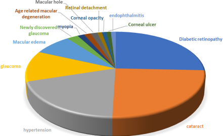
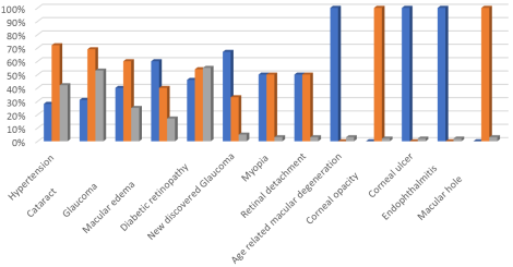
Background: Diabetes mellitus (DM) represents a significant global health issue, encompassing a range of heterogeneous disorders characterized by elevated blood glucose levels. Diabetes is classified into two types: type 1 (T1DM) and type 2 (T2DM). Diagnosis primarily relies on fasting blood glucose and HbA1c levels. Investigating the causes of vision impairment in diabetes is crucial for reducing the rates of blindness. Therefore, implementing screening for early diagnosis is essential to delay complications. Methodology: This study is a prospective descriptive analysis conducted from May 15, 2025, to October 20, 2025. The study was conducted at Doctor Khalil Ophthalmology Clinic in El-Obeid, North Kordofan, Sudan. This study included all individuals with diabetes attending the clinic for follow-up (520 patients). We assessed their visual acuity and selected all patients with poor vision uncorrected by glasses or other means of correction. Results: This study analysed 120 diabetic patients with impaired vision, comprising 58% females and 42% males. The most common condition identified was diabetic retinopathy, affecting 55% of participants, followed by cataract at 54%, hypertension at 46%, glaucoma at 25%, and macular oedema at 17%. Three percent of the cases had newly discovered glaucoma and myopia, and three patients had macular holes. Retinal detachments, corneal ulcers, corneal opacities, and endophthalmitis were also observed. Conclusion: Vision impairment associated with diabetes is prevalent in Sudan, largely attributable to ongoing conflict, which has resulted in inadequate health services and a scarcity of medications for glycaemic control in various regions.
| Published in | International Journal of Diabetes and Endocrinology (Volume 11, Issue 1) |
| DOI | 10.11648/j.ijde.20261101.11 |
| Page(s) | 1-6 |
| Creative Commons |
This is an Open Access article, distributed under the terms of the Creative Commons Attribution 4.0 International License (http://creativecommons.org/licenses/by/4.0/), which permits unrestricted use, distribution and reproduction in any medium or format, provided the original work is properly cited. |
| Copyright |
Copyright © The Author(s), 2026. Published by Science Publishing Group |
Diabetes Mellitus, Diabetic Retinopathy, Retinal Detachment, Macular Edema, Sudan War
≤ 59 Years | 60-64 | 65-70 | >70 | Total | ||
|---|---|---|---|---|---|---|
sex | Males | 14 | 8 | 16 | 12 | 50 |
Females | 12 | 18 | 22 | 18 | 70 | |
Total | 26 | 26 | 38 | 30 | 120 | |
Age group | 6l24 | 6l36 | 6l60 | <6l60 | Total |
|---|---|---|---|---|---|
≤ 59 Years | 0 | 4 | 6 | 16 | 26 |
60-64 | 2 | 2 | 8 | 14 | 26 |
65-70 | 8 | 2 | 8 | 20 | 38 |
>70 | 4 | 4 | 4 | 18 | 30 |
Total | 14 | 12 | 26 | 68 | 120 |
Variable | Males | Females | Total |
|---|---|---|---|
Diabetic retinopathy | 30 | 36 | 66 |
Cataract | 20 | 44 | 64 |
Hypertension | 14 | 36 | 50 |
Glaucoma | 12 | 18 | 30 |
Macular edema | 12 | 8 | 20 |
Newly discovered glaucoma | 4 | 2 | 6 |
Myopia | 2 | 2 | 4 |
Retinal detachment | 2 | 2 | 4 |
Age related macular degeneration | 4 | 0 | 4 |
Macular hole | 0 | 3 | 3 |
Corneal opacity | 0 | 2 | 2 |
Corneal ulcer | 2 | 0 | 2 |
Endophthalmitis | 2 | 0 | 2 |
Category | <59 years | 60-64 years | 65-70 years | >70 years | Total |
|---|---|---|---|---|---|
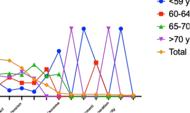
Diabetic retinopathy | 14 | 10 | 22 | 20 | 66 |
Cataract | 6 | 18 | 22 | 18 | 64 |
Hypertension | 6 | 10 | 16 | 18 | 50 |
Glaucoma | 2 | 6 | 14 | 8 | 30 |
Macular edema | 6 | 8 | 6 | 0 | 20 |
Newly discovered glaucoma | 4 | 0 | 2 | 0 | 6 |
myopia | 4 | 0 | 0 | 0 | 4 |
retinal detachment | 2 | 2 | 0 | 0 | 4 |
Age related macular degeneration | 0 | 0 | 0 | 4 | 4 |
Macular hole | 0 | 0 | 0 | 3 | 3 |
Corneal opacity | 2 | 0 | 0 | 0 | 2 |
Corneal ulcer | 2 | 0 | 0 | 0 | 2 |
Endophthalmitis | 0 | 0 | 0 | 2 | 2 |
DM | Diabetes Miletus |
T1DM | Type 1 DM |
T2DM | Type 2 DM |
| [1] |
Harreiter J, Roden M. Diabetes mellitus – Definition, Klassifikation, Diagnose, Screening und Prävention (Update 2023) [Diabetes mellitus: definition, classification, diagnosis, screening and prevention (Update 2023)]. Wien Klin Wochenschr. 2023 Jan; 135(Suppl 1): 7-17. German.
https://doi.org/10.1007/s00508-022-02122-y Epub 2023 Apr 20. |
| [2] | GBD 2019 Blindness and Vision Impairment Collaborators; Vision Loss Expert Group of the Global Burden of Disease Study. Causes of blindness and vision impairment in 2020 and trends over 30 years, and prevalence of avoidable blindness in relation to VISION 2020: the Right to Sight: an analysis for the Global Burden of Disease Study. Lancet Glob Health. 2021 Feb; 9(2): e144-e160. |
| [3] |
Lin KY, Hsih WH, Lin YB, Wen CY, Chang TJ. Update in the epidemiology, risk factors, screening, and treatment of diabetic retinopathy. J Diabetes Investig. 2021 Aug; 12(8): 1322-1325.
https://doi.org/10.1111/jdi.13480 Epub 2021 Jan 14. |
| [4] | Zarei-Ghanavati S, Hadi Y, Habibi A, Ashraf Khorasani M, Yoo SH. Cataract and diabetes: review of the literature. J Cataract Refract Surg. 2024 Dec 1; 50(12): 1275-1283. |
| [5] | Abumhadi M, Amin O, Mohammed S, Mohamedelkhair A, Osman E, Issak M, Mohammed W, Abdelmajid O, Abdoun A, Abdelaziz M. Knowledge, attitudes, and practices regarding diabetic retinopathy among patients with diabetes in Dongola, Northern State, Sudan, 2022: a cross-sectional study. Pan Afr Med J. 2024 Jun 28; 48: 77. |
| [6] |
Teo ZL, Tham YC, Yu M, Chee ML, Rim TH, Cheung N, Bikbov MM, Wang YX, Tang Y, Lu Y, Wong IY, Ting DSW, Tan GSW, Jonas JB, Sabanayagam C, Wong TY, Cheng CY. Global Prevalence of Diabetic Retinopathy and Projection of Burden through 2045: Systematic Review and Meta-analysis. Ophthalmology. 2021 Nov; 128(11): 1580-1591.
https://doi.org/10.1016/j.ophtha.2021.04.027 Epub 2021 May 1. |
| [7] |
Jia G, Sowers JR. Hypertension in Diabetes: An Update of Basic Mechanisms and Clinical Disease. Hypertension. 2021 Nov; 78(5): 1197-1205.
https://doi.org/10.1161/HYPERTENSIONAHA.121.17981 Epub 2021 Oct 4. |
| [8] | Sung JY, Lee KH, Jun JH, Lee MW. Changes in peripapillary microvasculature in patients with type 2 diabetes patients: effect of systemic hypertension. Sci Rep. 2023 Nov 9; 13(1): 19459. |
| [9] | Li Y, Mitchell W, Elze T, Zebardast N. Association Between Diabetes, Diabetic Retinopathy, and Glaucoma. Curr Diab Rep. 2021 Sep 8; 21(10): 38. |
| [10] | AlDarrab A, Al Jarallah OJ, Al Balawi HB. Association of diabetes, fasting glucose, and the risk of glaucoma: a systematic review and meta-analysis. Eur Rev Med Pharmacol Sci. 2023 Mar; 27(6): 2419-2427. |
| [11] |
Bandello F, Battaglia Parodi M, Lanzetta P, Loewenstein A, Massin P, Menchini F, Veritti D. Diabetic Macular Edema. Dev Ophthalmol. 2017; 58: 102-138.
https://doi.org/10.1159/000455277 Epub 2017 Mar 28. |
| [12] |
Yan A, Jones C, Demirel S, Chhablani J. Diabetic macular edema: Upcoming therapies. Graefes Arch Clin Exp Ophthalmol. 2025 Feb; 263(2): 249-258.
https://doi.org/10.1007/s00417-024-06595-7 Epub 2024 Jul 29. |
| [13] | Peled A, Raz I, Zucker I, Derazne E, Megreli J, Pinhas-Hamiel O, Einan-Lifshitz A, Morad Y, Pras E, Lutski M, Cukierman-Yaffe T, Mosenzon O, Tzur D, Tirosh A, Gerstein HC, Afek A, Twig G. Myopia and Early-Onset Type 2 Diabetes: A Nationwide Cohort Study. J Clin Endocrinol Metab. 2022 Jan 18; 107(2): e663-e671. |
| [14] | Lin JB, Narayanan R, Philippakis E, Yonekawa Y, Apte RS. Retinal detachment. Nat Rev Dis Primers. 2024 Mar 14; 10(1): 18. |
APA Style
Ibraheim, K. A. (2026). Factors Interrelated to Declining Vision Among Diabetic Patients in Sudan. International Journal of Diabetes and Endocrinology, 11(1), 1-6. https://doi.org/10.11648/j.ijde.20261101.11
ACS Style
Ibraheim, K. A. Factors Interrelated to Declining Vision Among Diabetic Patients in Sudan. Int. J. Diabetes Endocrinol. 2026, 11(1), 1-6. doi: 10.11648/j.ijde.20261101.11
AMA Style
Ibraheim KA. Factors Interrelated to Declining Vision Among Diabetic Patients in Sudan. Int J Diabetes Endocrinol. 2026;11(1):1-6. doi: 10.11648/j.ijde.20261101.11
@article{10.11648/j.ijde.20261101.11,
  author = {Khalil Ali Ibraheim},
  title = {Factors Interrelated to Declining Vision Among Diabetic Patients in Sudan},
  journal = {International Journal of Diabetes and Endocrinology},
  volume = {11},
  number = {1},
  pages = {1-6},
  doi = {10.11648/j.ijde.20261101.11},
  url = {https://doi.org/10.11648/j.ijde.20261101.11},
  eprint = {https://article.sciencepublishinggroup.com/pdf/10.11648.j.ijde.20261101.11},
  abstract = {Background: Diabetes mellitus (DM) represents a significant global health issue, encompassing a range of heterogeneous disorders characterized by elevated blood glucose levels. Diabetes is classified into two types: type 1 (T1DM) and type 2 (T2DM). Diagnosis primarily relies on fasting blood glucose and HbA1c levels. Investigating the causes of vision impairment in diabetes is crucial for reducing the rates of blindness. Therefore, implementing screening for early diagnosis is essential to delay complications. Methodology: This study is a prospective descriptive analysis conducted from May 15, 2025, to October 20, 2025. The study was conducted at Doctor Khalil Ophthalmology Clinic in El-Obeid, North Kordofan, Sudan. This study included all individuals with diabetes attending the clinic for follow-up (520 patients). We assessed their visual acuity and selected all patients with poor vision uncorrected by glasses or other means of correction. Results: This study analysed 120 diabetic patients with impaired vision, comprising 58% females and 42% males. The most common condition identified was diabetic retinopathy, affecting 55% of participants, followed by cataract at 54%, hypertension at 46%, glaucoma at 25%, and macular oedema at 17%. Three percent of the cases had newly discovered glaucoma and myopia, and three patients had macular holes. Retinal detachments, corneal ulcers, corneal opacities, and endophthalmitis were also observed. Conclusion: Vision impairment associated with diabetes is prevalent in Sudan, largely attributable to ongoing conflict, which has resulted in inadequate health services and a scarcity of medications for glycaemic control in various regions.},
 year = {2026}
}
TY  - JOUR
T1  - Factors Interrelated to Declining Vision Among Diabetic Patients in Sudan
AU  - Khalil Ali Ibraheim
Y1  - 2026/01/07
PY  - 2026
N1  - https://doi.org/10.11648/j.ijde.20261101.11
DO  - 10.11648/j.ijde.20261101.11
T2  - International Journal of Diabetes and Endocrinology
JF  - International Journal of Diabetes and Endocrinology
JO  - International Journal of Diabetes and Endocrinology
SP  - 1
EP  - 6
PB  - Science Publishing Group
SN  - 2640-1371
UR  - https://doi.org/10.11648/j.ijde.20261101.11
AB  - Background: Diabetes mellitus (DM) represents a significant global health issue, encompassing a range of heterogeneous disorders characterized by elevated blood glucose levels. Diabetes is classified into two types: type 1 (T1DM) and type 2 (T2DM). Diagnosis primarily relies on fasting blood glucose and HbA1c levels. Investigating the causes of vision impairment in diabetes is crucial for reducing the rates of blindness. Therefore, implementing screening for early diagnosis is essential to delay complications. Methodology: This study is a prospective descriptive analysis conducted from May 15, 2025, to October 20, 2025. The study was conducted at Doctor Khalil Ophthalmology Clinic in El-Obeid, North Kordofan, Sudan. This study included all individuals with diabetes attending the clinic for follow-up (520 patients). We assessed their visual acuity and selected all patients with poor vision uncorrected by glasses or other means of correction. Results: This study analysed 120 diabetic patients with impaired vision, comprising 58% females and 42% males. The most common condition identified was diabetic retinopathy, affecting 55% of participants, followed by cataract at 54%, hypertension at 46%, glaucoma at 25%, and macular oedema at 17%. Three percent of the cases had newly discovered glaucoma and myopia, and three patients had macular holes. Retinal detachments, corneal ulcers, corneal opacities, and endophthalmitis were also observed. Conclusion: Vision impairment associated with diabetes is prevalent in Sudan, largely attributable to ongoing conflict, which has resulted in inadequate health services and a scarcity of medications for glycaemic control in various regions.
VL  - 11
IS  - 1
ER  -
Dr. Khalil Ophthalmology Center, El-Obeid, Sudan;Department of Surgery, University of Kordofan, El-Obeid, Sudan;Department of Ophthalmology, El-Obeid Teaching Hospital, El-Obeid, Sudan;Sheikan College, El-Obeid, Sudan
Information