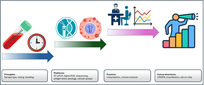
Accurate and timely viral diagnostics are central to modern clinical care and public health surveillance, guiding patient management, outbreak control, and population-level interventions. Even though advanced molecular technologies such as RT-qPCR, next-generation sequencing, and CRISPR-based assays have transformed viral detection, the diagnostic performance is shaped not only by analytical platforms but by the integrated flow of principles, platforms, and practice (3P framework). In essence, specimen type, timing of collection, transport conditions, and storage critically influence diagnostic sensitivity, which accounts for up to 60-70% of errors before laboratory analysis even begins. Direct detection approaches, including RT-qPCR, digital PCR, sequencing, and antigen assays, are examined as complementary tools rather than competing technologies, each occupying specialized clinical and public health niches. Indirect detection through serological and cellular immune assays is reviewed as a means of assessing exposure, immunity, and population-level transmission. The practical application of diagnostics is further discussed in key clinical contexts, including acute respiratory infections, chronic viral diseases, and arboviral illnesses, highlighting the importance of algorithmic testing strategies and epidemiological context. The real-world interpretation challenges are addressed, that emphasize on the pretest probability, false-positive and false-negative risks. Limitations of current evidence, including variability in study design, lack of standardization, and underrepresentation of low-resource settings, are critically assessed. Finally, emerging technologies such as CRISPR diagnostics, microfluidics, and lab-on-chip platforms are discussed as drivers of decentralized, rapid, and globally accessible testing. When biological principles, diagnostic technologies, and real-world clinical practice are considered together, it becomes clear that the true effectiveness of viral diagnostics does not rest on analytical performance alone. Rather, meaningful impact depends on how well diagnostic tools are integrated into everyday clinical decision making and public health systems. Looking ahead, the greatest advances are likely to come from diagnostic ecosystems that combine rapid detection with real-time data sharing and context-aware interpretation. Such an interconnected approach has the potential to transform viral diagnostics from isolated laboratory tests into continuous safeguards for both individual patients and population health. This review synthesizes current evidence across all stages of viral diagnostics, with particular emphasis on the often-overlooked pre-analytical and interpretative phases that dominate real world diagnostic error.
| Published in | International Journal of Infectious Diseases and Therapy (Volume 10, Issue 4) |
| DOI | 10.11648/j.ijidt.20251004.13 |
| Page(s) | 93-104 |
| Creative Commons |
This is an Open Access article, distributed under the terms of the Creative Commons Attribution 4.0 International License (http://creativecommons.org/licenses/by/4.0/), which permits unrestricted use, distribution and reproduction in any medium or format, provided the original work is properly cited. |
| Copyright |
Copyright © The Author(s), 2025. Published by Science Publishing Group |
Viral Infections, Viral Diagnostics, RT-PCR, Serology
Domain | Sub-category | Key methods / examples | Strengths | Limitations | Clinical contexts |
|---|---|---|---|---|---|
Principles (pre-analytical) | Specimen type | NP/OP swabs, saliva, blood, stool, CSF, biopsies | High sensitivity if optimal sample chosen | Invasive procedures, degradation risk | Respiratory viruses (swabs), systemic viruses (blood), neurotropic viruses (CSF) |
Timing | Early sampling (3-5 days post symptoms for SARS-CoV-2) | Maximizes viral load detection | Late collection leading to false negatives | Acute infections | |
Handling and transport | Viral transport medium, stabilization buffers | Preserves nucleic acids/proteins | Cold chain needed, freeze-thaw damage | All infections | |
Platforms (analytical) | Nucleic acid amplification | RT-qPCR, digital PCR | High sensitivity, quantification | Cost, contamination risk | SARS-CoV-2, HIV, HBV |
Sequencing | NGS, metagenomics | Pathogen discovery, surveillance | Expensive, infrastructure heavy | Novel pathogens, genomic epidemiology | |
Antigen detection | Rapid antigen, CLIA, ECLIA | Fast, scalable | Lower sensitivity, time-window limited | Respiratory viruses, mass screening | |
Serology | ELISA, neutralization assays | Detects past exposure, population immunity | Cross-reactivity, waning antibodies | Dengue vs Zika, COVID-19 serosurveys | |
Cellular assays | ELISpot, IGRA | Detects T cell immunity | Complex, limited standardization | SARS-CoV-2 | |
Practice (interpretation and application) | Acute infections | Algorithmic testing (Antigen and PCR) | Rapid triage, confirmatory testing | False negatives if misapplied | RSV, COVID-19 |
Chronic infections | Viral load, resistance testing | Guides therapy, monitors response | Requires infrastructure | HIV, HBV, HCV | |
Arboviruses | Serology and molecular antigens | Holistic diagnosis | Cross-reactivity common | Dengue, Zika | |
Real-world interpretation | Bayesian reasoning, pretest probability | Integrates epidemiology and test data | Requires clinician training | All infections |
Technique | Sensitivity | Strengths | Weaknesses |
|---|---|---|---|
RT-qPCR | High | Gold standard; high specificity and sensitivity; rapid turnaround; widely available | Requires cold chain and technical expertise; contamination risk; semi-quantitative (Ct values vary) |
Digital PCR | Very high | Absolute quantification; excellent for low-copy detection (early HIV, HBV, residual SARS-CoV-2); reproducible | Expensive; limited availability; lower throughput compared to RT-qPCR |
NGS | High (context-dependent) | Unbiased detection; pathogen discovery; genomic surveillance; detects co-infections and resistance mutations | Costly; requires bioinformatics and infrastructure; clinical interpretation can be complex |
Rapid Antigen Tests (Lateral flow) | Low to moderate | Fast (<30 min); inexpensive; point-of-care; scalable for mass screening | Lower sensitivity (especially late infection); performance depends on viral load and timing; more false negatives |
Laboratory-based Antigen Assays (CLIA, ECLIA) | Moderate to high | High-throughput; automated; more sensitive than lateral flow | Still less sensitive than PCR; may detect non-viable viruses; requires lab setting |
Serology (ELISA, Neutralization assays) | Moderate | Detects past exposure and immunity; useful for surveillance; neutralization assays show functional protection | Cross-reactivity (flaviviruses); waning antibodies; cannot distinguish infection vs vaccination easily |
Cellular assays (ELISpot, IGRA) | Variable (low-moderate) | Captures T cell immunity; longer-lasting responses than antibodies; adds depth to immune profiling | Technically demanding; requires fresh samples; limited standardization; interpretation still evolving |
3P | Principles, Platforms, and Practice |
Cas12 | CRISPR-associated Protein 12 |
Cas13 | CRISPR-associated Protein 13 |
CLIA | Chemiluminescent Immunoassay |
COVID-19 | Coronavirus Disease 2019 |
CRISPR | Clustered Regularly Interspaced Short Palindromic Repeats |
Ct | Cycle Threshold |
EC | External Control |
ECLIA | Electrochemiluminescence Immunoassay |
ELISA | Enzyme-Linked Immunosorbent Assay |
ELISpot | Enzyme-Linked ImmunoSpot |
HBV | Hepatitis B Virus |
HCV | Hepatitis C Virus |
HIV | Human Immunodeficiency Virus |
IC | Internal Control |
IgA | Immunoglobulin A |
IgG | Immunoglobulin G |
IgM | Immunoglobulin M |
IGRA | Interferon-Gamma Release Assay |
NGS | Next-Generation Sequencing |
PCR | Polymerase Chain Reaction |
RNA | Ribonucleic Acid |
RSV | Respiratory Syncytial Virus |
RT-PCR | Reverse Transcription Polymerase Chain Reaction |
RT-qPCR | Reverse Transcription Quantitative Polymerase Chain Reaction |
SARS-CoV-2 | Severe Acute Respiratory Syndrome Coronavirus 2 |
SHERLOCK | Specific High-sensitivity Enzymatic Reporter unLOCKing |
DETECTR | DNA Endonuclease Targeted CRISPR Trans Reporter |
UDG | Uracil-DNA Glycosylase |
| [1] | Dronina, J., U. Samukaite-Bubniene, and A. Ramanavicius, Advances and insights in the diagnosis of viral infections. Journal of Nanobiotechnology, 2021. 19(1): p. 348. |
| [2] | Misra, A. and E. A. Powell, Preanalytical Challenges of Molecular Microbiology Tests. Clinics in Laboratory Medicine, 2024. 44(1): p. 33–43. |
| [3] | Pallen, M. J., N. J. Loman, and C. W. Penn, High-throughput sequencing and clinical microbiology: progress, opportunities and challenges. Current opinion in microbiology, 2010. 13(5): p. 625–631. |
| [4] | Fournier, P. E., et al., Modern clinical microbiology: new challenges and solutions. Nature Reviews Microbiology, 2013. 11(8): p. 574–585. |
| [5] | Lippi, G., et al., Causes, consequences, detection, and prevention of identification errors in laboratory diagnostics. Clinical chemistry and laboratory medicine, 2009. 47(2): p. 143–153. |
| [6] | van Zyl, G., et al., Lessons in diagnostic virology: expected and unexpected sources of error. Reviews in medical virology, 2019. 29(4): p. e2052. |
| [7] | Chavez, V., Sources of pre-analytical, analytical and post-analytical errors in the microbiology laboratory, in Accurate results in the clinical laboratory. 2019, Elsevier. p. 377–384. |
| [8] | Hardt, M., et al., Pre-analytical sample stabilization by different sampling devices for PCR-based COVID-19 diagnostics. New Biotechnology, 2022. 70: p. 19–27. |
| [9] | Rasool, G., et al., COVID-19: Clinical laboratory diagnosis and monitoring of novel coronavirus infected patients using molecular, serological and biochemical markers: A review. International journal of immunopathology and pharmacology, 2022. 36: p. 03946320221115316. |
| [10] | Padalko, E., et al., Preanalytical variables influencing the interpretation and reporting of biological tests on blood samples of living and deceased donors for human body materials. Cell and tissue banking, 2024. 25(2): p. 509–520. |
| [11] | Singh, S., A. Yadav, and S. Sharma, Choice of Samples for Molecular Diagnosis of Viral Disease, in Molecular Diagnostics for Viral Diseases: Challenges and Emerging Concepts. 2025, Springer. p. 47–63. |
| [12] | Liu, B. M., et al., Laboratory diagnosis of CNS infections in children due to emerging and re-emerging neurotropic viruses. Pediatric research, 2024. 95(2): p. 543–550. |
| [13] | Olie, S. E., et al., Molecular diagnostics in cerebrospinal fluid for the diagnosis of central nervous system infections. Clinical Microbiology Reviews, 2024. 37(4): p. e00021–24. |
| [14] | Basso, D., et al., SARS-CoV-2 RNA identification in nasopharyngeal swabs: issues in pre-analytics. Clinical Chemistry and Laboratory Medicine (CCLM), 2020. 58(9): p. 1579–1586. |
| [15] | Lee, C. Y. P., et al., Serological approaches for COVID-19: epidemiologic perspective on surveillance and control. Frontiers in immunology, 2020. 11: p. 879. |
| [16] | Hardt, M., et al., Pre-analytical properties of different respiratory viruses for PCR-based detection: Comparative analysis of sampling devices and sample stabilization solutions. New Biotechnology, 2024. 79: p. 60–70. |
| [17] | Ilardo, C., et al., Performance and pre-analytical stability of self-collected samples versus clinician cervical samples for the detection of HPV16, HPV18 and a pool of 12 other HPV types on the Roche Cobas 8800 System. New Microbiol, 2022. 45(2): p. 111–114. |
| [18] | Setzer, M., J. Juusola, and J. Ballantyne, Recovery and stability of RNA in vaginal swabs and blood, semen, and saliva stains. Journal of forensic sciences, 2008. 53(2): p. 296–305. |
| [19] | Zoch-Lesniak, B., et al., The Respiratory Specimen Collection Trial (ReSpeCT): a randomized controlled trial to compare quality and timeliness of respiratory sample collection in the home by parents and healthcare workers from children aged< 2 years. Journal of the Pediatric Infectious Diseases Society, 2020. 9(2): p. 134–141. |
| [20] | Dunn, J. J., Specimen collection, transport, and processing: virology. Manual of clinical microbiology, 2015: p. 1405–1421. |
| [21] | Busby, E. J., Metrology and Molecular Diagnosis of Infection. 2020, UCL (University College London). |
| [22] | Ji, X., et al., The impact of repeated freeze–thaw cycles on the quality of biomolecules in four different tissues. Biopreservation and biobanking, 2017. 15(5): p. 475–483. |
| [23] | Abulfathi, F. A., Evaluation and validation of room temperature biospecimens transportation and storage technologies as an alternative cost effective solution to cold chain logistics and storage within biobanking and/or diagnostics. 2017, Stellenbosch: Stellenbosch University. |
| [24] | Barranco, T., et al., Changes of salivary biomarkers under different storage conditions: effects of temperature and length of storage. Biochem Med (Zagreb), 2019. 29(1): p. 010706. |
| [25] | Lustrek, M., et al., Influenza A, Influenza B, human respiratory syncytial virus and SARSCoV-2 molecular diagnostics and epidemiology in the post COVID-19 era. Respiratory Research, 2024. 25(1): p. 234. |
| [26] | Vester, D., et al., Real-time RT-qPCR assay for the analysis of human influenza A virus transcription and replication dynamics. Journal of virological methods, 2010. 168(1-2): p. 63–71. |
| [27] | Pereira, L., et al., Standardization of a high-performance RT-qPCR for viral load absolute quantification of influenza A. Journal of Virological Methods, 2022. 301: p. 114439. |
| [28] | Watzinger, F., K. Ebner, and T. Lion, Detection and monitoring of virus infections by real-time PCR. Molecular aspects of medicine, 2006. 27(2-3): p. 254–298. |
| [29] | Reta, D. H., et al., Molecular and immunological diagnostic techniques of medical viruses. International Journal of Microbiology, 2020. 2020(1): p. 8832728. |
| [30] | Chauhan, N., S. Soni, and U. Jain, Optimizing testing regimes for the detection of COVID-19 in children and older adults. Expert review of molecular diagnostics, 2021. 21(10): p. 999–1016. |
| [31] | Merino, I., et al., Digital PCR applications for the diagnosis and management of infection in critical care medicine. Critical Care, 2022. 26(1): p. 63. |
| [32] | Meng, Z., et al., Applications of laboratory findings in the prevention, diagnosis, treatment, and monitoring of COVID-19. Signal transduction and targeted therapy, 2021. 6(1): p. 316. |
| [33] | Shaffaf, T., COVID-9 Detection Strategies: Recent Advances and Future Prospects. 2021. |
| [34] | Lambisia, A. W., Comparison of the Diagnostic Performance of TaqMan Array Cards, Enzyme Immunoassay, Real-Time PCR and Next Generation Sequencing in Investigation of Five Common Diarrhoea-Associated Enteric Viruses in Kilifi, Kenya. 2021, JKUAT-COHES. |
| [35] | Yuan, X., et al., Current and perspective diagnostic techniques for COVID-19. ACS infectious diseases, 2020. 6(8): p. 1998–2016. |
| [36] | Nguyen, N. N., et al., Development of diagnostic tests for detection of SARS-CoV-2. Diagnostics, 2020. 10(11): p. 905. |
| [37] | Iliescu, F. S., et al., Point-of-care testing—the key in the battle against SARS-CoV-2 pandemic. Micromachines, 2021. 12(12): p. 1464. |
| [38] | Pandey, S. K., et al., Diagnostic tools for rapid screening and detection of SARS-CoV-2 infection. Vaccines, 2022. 10(8): p. 1200. |
| [39] | Gómez‐Vicente, E., et al., Comparative evaluation of chemiluminescent immunoassay and enzyme‐linked immunosorbent assays for the diagnosis of West Nile virus infections. APMIS, 2022. 130(4): p. 215–220. |
| [40] | Wadood, M. and M. Usman, Comparative analysis of electrochemiluminescence assay and chemiluminescent microparticle immunoassay for the screening of hepatitis C. Indian Journal of Hematology and Blood Transfusion, 2019. 35(1): p. 131–136. |
| [41] | Lei, Y., et al., Distribution characteristics of SARS-CoV-2 IgM/IgG in false-positive results detected by chemiluminescent immunoassay. Open Life Sciences, 2022. 17(1): p. 1487–1496. |
| [42] | Nicolai, E., et al., The evolution of serological assays during two years of the COVID-19 pandemic: from an easy-to-use screening tool for identifying current infections to laboratory algorithms for discovering immune protection and optimizing vaccine administration. COVID, 2024. 4(8): p. 1272–1290. |
| [43] | Boonham, N., et al., Methods in virus diagnostics: from ELISA to next generation sequencing. Virus research, 2014. 186: p. 20–31. |
| [44] | Lochmanová, A., et al., Comparison of two commercially available interferon-γ release assays for T-cell-mediated immunity and evaluation of humoral immunity against SARS-CoV-2 in healthcare workers. Diagnostics, 2023. 13(4): p. 637. |
| [45] | Dan, J. M., et al., Immunological memory to SARS-CoV-2 assessed for up to 8 months after infection. Science, 2021. 371(6529): p. eabf4063. |
| [46] | Sette, A. and S. Crotty, Adaptive immunity to SARS-CoV-2 and COVID-19. Cell, 2021. 184(4): p. 861–880. |
| [47] | Jacobs, A., The role of antibodies in tuberculosis. 2024. |
| [48] | Sakshi, et al., Novel diagnostic methods for emerging respiratory viral infection, in Emerging Human Viral Diseases, Volume I: Respiratory and Haemorrhagic Fever. 2023, Springer. p. 565–585. |
| [49] | Saha, A., et al., Beyond the Pandemic Era: Recent Advances and Efficacy of SARS-CoV-2 Vaccines Against Emerging Variants of Concern. Vaccines, 2025. 13(4): p. 424. |
| [50] | Berger, A., W. Preiser, and H. W. Doerr, The role of viral load determination for the management of human immunodeficiency virus, hepatitis B virus and hepatitis C virus infection. Journal of clinical virology, 2001. 20(1-2): p. 23–30. |
| [51] | Martinello, M., et al., Management of acute HCV infection in the era of direct-acting antiviral therapy. Nature reviews Gastroenterology & hepatology, 2018. 15(7): p. 412–424. |
| [52] | Shaw, T., A. Bartholomeusz, and S. Locarnini, HBV drug resistance: mechanisms, detection and interpretation. Journal of hepatology, 2006. 44(3): p. 593–606. |
| [53] | Gomes da Silva, P., et al., Serological cross-reactivity in zoonotic flaviviral infections of medical importance. Antibodies, 2023. 12(1): p. 18. |
| [54] | Chan, K. R., et al., Serological cross-reactivity among common flaviviruses. Frontiers in Cellular and Infection Microbiology, 2022. 12: p. 975398. |
| [55] | Dragoni, F., Antiviral drug development for treatment of acute and chronic viral infections. 2021. |
| [56] | Niesters, H., Molecular and diagnostic clinical virology in real time. Clinical microbiology and infection, 2004. 10(1): p. 5–11. |
| [57] | Vainionpää, R., M. Waris, and P. Leinikki, Diagnostic techniques: serological and molecular approaches. Reference module in biomedical sciences, 2015: p. B978–0–12–801238–3.02558–7. |
| [58] | Bentley, P. M., Error rates in SARS-CoV-2 testing examined with Bayesian inference. medRxiv, 2020: p. 2020.12. 17.20248402. |
| [59] | Wu, Y., et al., Bridging the gaps in test interpretation of SARS-CoV-2 through Bayesian network modelling. Epidemiology & Infection, 2021. 149: p. e166. |
| [60] | Klarkowski, D., et al., Causes of false-positive HIV rapid diagnostic test results. Expert review of anti-infective therapy, 2014. 12(1): p. 49–62. |
| [61] | Braunstein, G. D., et al., False positive results with SARS-CoV-2 RT-PCR tests and how to evaluate a RT-PCR-positive test for the possibility of a false positive result. Journal of Occupational and Environmental Medicine, 2021. 63(3): p. e159–e162. |
| [62] | Mitchell, S. L. and M. J. Loeffelholz, Considerations regarding interpretation of positive SARS-CoV-2 molecular results with late cycle threshold values. Journal of Clinical Microbiology, 2022. 60(9): p. e00501–22. |
| [63] | Morgan, D. J., et al., Accuracy of practitioner estimates of probability of diagnosis before and after testing. JAMA internal medicine, 2021. 181(6): p. 747–755. |
| [64] | Baduashvili, A., A. T. Evans, and T. Cutler, How to understand and teach P values: a diagnostic test framework. J Clin Epidemiol, 2020. 122: p. 49–55. |
| [65] | Jarrom, D., et al., Effectiveness of tests to detect the presence of SARS-CoV-2 virus, and antibodies to SARS-CoV-2, to inform COVID-19 diagnosis: a rapid systematic review. BMJ evidence-based medicine, 2022. 27(1): p. 33–45. |
| [66] | Tsang, N. N. Y., et al., Diagnostic performance of different sampling approaches for SARS-CoV-2 RT-PCR testing: a systematic review and meta-analysis. The Lancet Infectious Diseases, 2021. 21(9): p. 1233–1245. |
| [67] | Chou, R., P. Easterbrook, and M. Hellard, Methodological challenges in appraising evidence on diagnostic testing for WHO guidelines on hepatitis B and hepatitis C virus infection. BMC infectious diseases, 2017. 17(Suppl 1): p. 694. |
| [68] | Onwuchekwa, C., et al., Underascertainment of respiratory syncytial virus infection in adults due to diagnostic testing limitations: a systematic literature review and meta-analysis. The Journal of infectious diseases, 2023. 228(2): p. 173–184. |
| [69] | Kiselev, D., et al., Current trends in diagnostics of viral infections of unknown etiology. Viruses, 2020. 12(2): p. 211. |
| [70] | Van Zant, W. and P. Ray, Democratization of Point-of-Care Viral Biosensors: Bridging the Gap from Academia to the Clinic. Biosensors, 2025. 15(7): p. 436. |
| [71] | Sarantopoulos, A., et al., Artificial intelligence in infectious disease clinical practice: an overview of gaps, opportunities, and limitations. Tropical Medicine and Infectious Disease, 2024. 9(10): p. 228. |
| [72] | Baumfeld Andre, E., et al., The current landscape and emerging applications for real‐world data in diagnostics and clinical decision support and its impact on regulatory decision making. Clinical Pharmacology & Therapeutics, 2022. 112(6): p. 1172–1182. |
| [73] | Pai, N. P., et al., Barriers to implementation of rapid and point-of-care tests for human immunodeficiency virus infection: findings from a systematic review (1996–2014). Point of care, 2015. 14(3): p. 81–87. |
| [74] | Trickey, A., et al., Impact of hepatitis C virus point-of-care RNA viral load testing compared with laboratory-based testing on uptake of RNA testing and treatment, and turnaround times: a systematic review and meta-analysis. The lancet Gastroenterology & hepatology, 2023. 8(3): p. 253–270. |
| [75] | Wei, L., et al., CRISPR/Cas Multiplexed Biosensing: Advances, Challenges, and Perspectives. Analytical Chemistry, 2025. |
| [76] | Zu, Y., H. Chang, and Z. Cui, Molecular point-of-care testing technologies: current status and challenges. Nexus, 2025. |
| [77] | Wang, Y., et al., Advances in nucleic acid assays for infectious disease: the role of microfluidic technology. Molecules, 2024. 29(11): p. 2417. |
| [78] | Panwar, R., H. Churi, and S. Dave, Point-of-care electrochemical biosensors using CRISPR/Cas for RNA analysis, in Biosensors for Emerging and Re-Emerging Infectious Diseases. 2022, Elsevier. p. 317–333. |
APA Style
Albargy, H. (2025). 3P in Diagnostics of Viral Infections: Principles, Platforms, and Practice. International Journal of Infectious Diseases and Therapy, 10(4), 93-104. https://doi.org/10.11648/j.ijidt.20251004.13
ACS Style
Albargy, H. 3P in Diagnostics of Viral Infections: Principles, Platforms, and Practice. Int. J. Infect. Dis. Ther. 2025, 10(4), 93-104. doi: 10.11648/j.ijidt.20251004.13
AMA Style
Albargy H. 3P in Diagnostics of Viral Infections: Principles, Platforms, and Practice. Int J Infect Dis Ther. 2025;10(4):93-104. doi: 10.11648/j.ijidt.20251004.13
@article{10.11648/j.ijidt.20251004.13,
  author = {Hassan Albargy},
  title = {3P in Diagnostics of Viral Infections: Principles, Platforms, and Practice},
  journal = {International Journal of Infectious Diseases and Therapy},
  volume = {10},
  number = {4},
  pages = {93-104},
  doi = {10.11648/j.ijidt.20251004.13},
  url = {https://doi.org/10.11648/j.ijidt.20251004.13},
  eprint = {https://article.sciencepublishinggroup.com/pdf/10.11648.j.ijidt.20251004.13},
  abstract = {Accurate and timely viral diagnostics are central to modern clinical care and public health surveillance, guiding patient management, outbreak control, and population-level interventions. Even though advanced molecular technologies such as RT-qPCR, next-generation sequencing, and CRISPR-based assays have transformed viral detection, the diagnostic performance is shaped not only by analytical platforms but by the integrated flow of principles, platforms, and practice (3P framework). In essence, specimen type, timing of collection, transport conditions, and storage critically influence diagnostic sensitivity, which accounts for up to 60-70% of errors before laboratory analysis even begins. Direct detection approaches, including RT-qPCR, digital PCR, sequencing, and antigen assays, are examined as complementary tools rather than competing technologies, each occupying specialized clinical and public health niches. Indirect detection through serological and cellular immune assays is reviewed as a means of assessing exposure, immunity, and population-level transmission. The practical application of diagnostics is further discussed in key clinical contexts, including acute respiratory infections, chronic viral diseases, and arboviral illnesses, highlighting the importance of algorithmic testing strategies and epidemiological context. The real-world interpretation challenges are addressed, that emphasize on the pretest probability, false-positive and false-negative risks. Limitations of current evidence, including variability in study design, lack of standardization, and underrepresentation of low-resource settings, are critically assessed. Finally, emerging technologies such as CRISPR diagnostics, microfluidics, and lab-on-chip platforms are discussed as drivers of decentralized, rapid, and globally accessible testing. When biological principles, diagnostic technologies, and real-world clinical practice are considered together, it becomes clear that the true effectiveness of viral diagnostics does not rest on analytical performance alone. Rather, meaningful impact depends on how well diagnostic tools are integrated into everyday clinical decision making and public health systems. Looking ahead, the greatest advances are likely to come from diagnostic ecosystems that combine rapid detection with real-time data sharing and context-aware interpretation. Such an interconnected approach has the potential to transform viral diagnostics from isolated laboratory tests into continuous safeguards for both individual patients and population health. This review synthesizes current evidence across all stages of viral diagnostics, with particular emphasis on the often-overlooked pre-analytical and interpretative phases that dominate real world diagnostic error.},
 year = {2025}
}
TY  - JOUR
T1  - 3P in Diagnostics of Viral Infections: Principles, Platforms, and Practice
AU  - Hassan Albargy
Y1  - 2025/12/29
PY  - 2025
N1  - https://doi.org/10.11648/j.ijidt.20251004.13
DO  - 10.11648/j.ijidt.20251004.13
T2  - International Journal of Infectious Diseases and Therapy
JF  - International Journal of Infectious Diseases and Therapy
JO  - International Journal of Infectious Diseases and Therapy
SP  - 93
EP  - 104
PB  - Science Publishing Group
SN  - 2578-966X
UR  - https://doi.org/10.11648/j.ijidt.20251004.13
AB  - Accurate and timely viral diagnostics are central to modern clinical care and public health surveillance, guiding patient management, outbreak control, and population-level interventions. Even though advanced molecular technologies such as RT-qPCR, next-generation sequencing, and CRISPR-based assays have transformed viral detection, the diagnostic performance is shaped not only by analytical platforms but by the integrated flow of principles, platforms, and practice (3P framework). In essence, specimen type, timing of collection, transport conditions, and storage critically influence diagnostic sensitivity, which accounts for up to 60-70% of errors before laboratory analysis even begins. Direct detection approaches, including RT-qPCR, digital PCR, sequencing, and antigen assays, are examined as complementary tools rather than competing technologies, each occupying specialized clinical and public health niches. Indirect detection through serological and cellular immune assays is reviewed as a means of assessing exposure, immunity, and population-level transmission. The practical application of diagnostics is further discussed in key clinical contexts, including acute respiratory infections, chronic viral diseases, and arboviral illnesses, highlighting the importance of algorithmic testing strategies and epidemiological context. The real-world interpretation challenges are addressed, that emphasize on the pretest probability, false-positive and false-negative risks. Limitations of current evidence, including variability in study design, lack of standardization, and underrepresentation of low-resource settings, are critically assessed. Finally, emerging technologies such as CRISPR diagnostics, microfluidics, and lab-on-chip platforms are discussed as drivers of decentralized, rapid, and globally accessible testing. When biological principles, diagnostic technologies, and real-world clinical practice are considered together, it becomes clear that the true effectiveness of viral diagnostics does not rest on analytical performance alone. Rather, meaningful impact depends on how well diagnostic tools are integrated into everyday clinical decision making and public health systems. Looking ahead, the greatest advances are likely to come from diagnostic ecosystems that combine rapid detection with real-time data sharing and context-aware interpretation. Such an interconnected approach has the potential to transform viral diagnostics from isolated laboratory tests into continuous safeguards for both individual patients and population health. This review synthesizes current evidence across all stages of viral diagnostics, with particular emphasis on the often-overlooked pre-analytical and interpretative phases that dominate real world diagnostic error.
VL  - 10
IS  - 4
ER  -
Department of Medical Laboratories, Shaqra University, Shaqra, Saudi Arabia
Information