Stroke is a major public health problem worldwide, the third leading cause of death, and the leading cause of acquired physical disability in adults. Compared with cerebral infarctions, hemorrhagic strokes tend to occur earlier in life and have a poorer prognosis. We aim to study the computed tomography aspects of hemorrhagic strokes in the CHUR-OHG imaging department. This was a descriptive cross-sectional study with retrospective data collection, from 29 September 2022 to 31 August 2023. It concerning patients with hemorrhagic stroke confirmed by brain scan. The mean age of 58 included patients was 59 ± 15 years and the sex ratio was 1.23. Acquisition without injection of contrast medium was performed in 98.28% of cases. Intracerebral hemorrhage was the most frequent lesion (98.28%). It was isolated or associated with ventricular flooding (36.21%) or meningeal hemorrhage (12.07%). It was divided into capsular hematomas in 84.21% of cases with mainly lenticular and thalamic extensions in 49.12% and 36.84% of cases respectively. A mass effect was found in 43.10% of cases on the ventricles and 44.83% on the midline, which was deviated by an average of 6.75 ± 3.1 mm. Cerebral involvement and edema were present in 34.48% and 6.90% of cases respectively. Hemorrhagic strokes is a serious and fatal disease. Primary prevention focusing on risk factors, early diagnosis using Computed tomography or, at best, magnetic resonance imaging, and appropriate management are essential to improve the prognosis.
This is an Open Access article, distributed under the terms of the Creative Commons Attribution 4.0 International License (http://creativecommons.org/licenses/by/4.0/), which permits unrestricted use, distribution and reproduction in any medium or format, provided the original work is properly cited.
Stroke is a major public health problem worldwide, the third leading cause of death, and the leading cause of acquired physical disability in adults
[1]
Raveloson NE, Zodaly N, Rakotoarivony ST, Mbolamena RL, Randriamiarana JM. Aspects épidémiocliniques, évolutifs et tomodensitométriques des accidents vasculaires cérébraux hémorragiques (34 cas). [Epidemiological, evolutionary, and computed tomography aspects of haemorrhagic stroke (34 cases)]. Revue d’anesthésie-réanimation et de médecine d’urgence 2011; 3(1): 15–9.
[1]
. Compared with cerebral infarctions, hemorrhagic strokes tend to occur earlier in life and have a poorer prognosis, with a risk of death in the first year in around 50% of cases
[2]
Lecmlerc X, Khalil C, Silvera S, Gauvrit Y, Bracard S, Meder F, Pruvo P. Imagerie des hématomes intracérébraux non traumatiques. [Imaging of non-traumatic intracerebral haematomas]. Journal of Neuroradiologie 2016.
. They account for 20% of strokes; however, they are responsible for 30 to 50% of deaths
[3]
Broderick JP, Adams HP, Barsan W, Feinberg W, Feldmann E, Grotta J, et al. Guidelines for the Management of Spontaneous Intracerebral Hemorrhage: A statement for healthcare professionals from a special writing group of the Stroke Council, American Heart Association.. Stroke 1999; 30(4): 905-15.
Amarenco P. Attaque cérébrale: qu’est-ce qu’une hémorragie cérébrale, une hémorragie méningée? [Stroke: what is a cerebral hemorrhage, a meningeal hemorrhage?] Corresondances en neurologie vasculaire 2003; 3: 11-15.
[4]
. The stroke was diagnosed solely on clinical grounds. The discovery of computed tomography (CT) by Godfrey Newbold Hounsfield and its introduction into medical practice in 1970 revolutionized this diagnostic approach and led to a new approach to patient examination. Likewise, Paul Castaigne points out it is more appropriate for making a fairly accurate diagnosis
[5]
Sonhaye L, Tchaou M, Adjenou K, Agoda-Koussema LK, N’Timon B, N’Dakena, K. Aspects scanographiques des accidents vasculaires cérébraux au Chu Campus de Lome, Togo (à propos de 314 cas). [Scanographical aspects of cerebral vascular accidents at the Chu campus of Lome, Togo (About 314 cases)]. J Rech Sci Univ Lomé (Togo), 2011: 31–6.
[5]
. CT is the basic examination in developed countries
[5]
Sonhaye L, Tchaou M, Adjenou K, Agoda-Koussema LK, N’Timon B, N’Dakena, K. Aspects scanographiques des accidents vasculaires cérébraux au Chu Campus de Lome, Togo (à propos de 314 cas). [Scanographical aspects of cerebral vascular accidents at the Chu campus of Lome, Togo (About 314 cases)]. J Rech Sci Univ Lomé (Togo), 2011: 31–6.
[5]
but has only recently been introduced (less than three years ago) at the Ouahigouya Regional University Hospital (CHUR-OHG). Given the seriousness and serious consequences of hemorrhagic strokes, we thought it appropriate to study the CT aspects of this pathology at CHUR-OHG.
2. Materials and Methods
This was a descriptive cross-sectional study with retrospective data collection. It was conducted in the Medical Imaging Department of the CHUR-OHG. It covered the period from 29 September 2022 to 31 August 2023.
The study included all patients whose cerebral CT scan performed for clinical suspicion of stroke confirmed an hemorrhagic strokes.
The data were collected from the CT images, the registry, and the reports of the CT examinations on a pre-established individual form. It included socio-demographic variables (age, sex), patient medical history, clinical data, the protocol used for the CT scan, types of hemorrhagic lesions, impact of the lesions, and etiologies of the lesions.
The data was analyzed using EPI info software version 7.2.5.
Patient anonymity was respected, and we obtained permission from the Director General of CHUR-OHG before the study began.
3. Results
3.1. General Information
During the study period (11 months), 9707 patients were admitted to the Medical Imaging Department and 58 were included in this study, giving a frequency of 0.60%.
The mean age was 59 (± 15) with the extremes of 11 and 103 years. The 45-64 years group accounted for 53.45% of cases. The sample comprised 55.17% men with a sex ratio of 1.23.
Arterial hypertension was present in 20.69% of patients. Breast cancer and nephro-angiosclerosis were present in one patient each.
The neurological signs that gave rise to a suspicion of stroke were represented by a motor deficit in 87.93% of patients, with a sudden onset in 55.17% of cases. The motor deficit affected the left hemisphere in 61% of cases and consisted of hemiplegia in 74.07%, aphasia in 37.04%, and facial paralysis in 11.11% of cases. Headache was also reported by 43% of patients, or associated with vertigo in 29%, altered consciousness in 18.97%, and dysarthria and convulsions in 14%.
3.2. CT Scan Data
The helical acquisition technique without injection of contrast medium was used in 98.28% of patients.
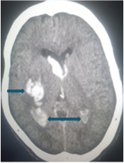
Figure 1. Cerebral CT scan without injection of contrast medium showing in axial section, at the parenchymal window, a large right capsulolenticular hematoma (→) with flooding of the lateral ventricles (↔).
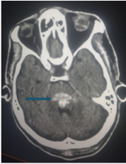
Figure 2. Cerebral CT scan without injection of contrast medium showing in axial section, at the parenchymal window, a recent hemorrhage of the annular protuberance of the brainstem (→).
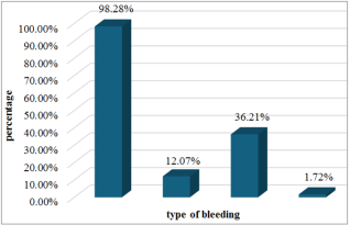
The intraparenchymal hematoma was found in 98.28% of cases, associated with subarachnoid hemorrhage in 12.07%, and ventricular flooding in 36.21% (figures 1 and 2). Only one patient had a subdural hematoma.
Figure 3 shows the distribution of patients by type of bleed.
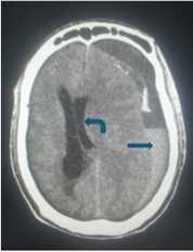
Figure 4. Cerebral CT scan without injection of contrast medium showing axial section at the parenchymal window a left frontoparietal subdural hematoma (→) with ipsilateral ventricular compression and subfalcorial involvement (←).
The intraparenchymal hematoma was preferentially capsular in 84.21%, lenticular in 49.12%, thalamic in 36.84%, pontine in 7.02%, caudate in 5.26%, truncular in 3.51%, cerebellar and subcallosal in 1.75% each.
There was a mass effect on the midline in 44.83% of cases and on the ventricles in 43.10% of cases. Cerebral involvement (figure 4) and edema were present in 34.48% and 6.90% of cases respectively.
Other associated lesions included sinusitis with 24%, cortico-subcortical atrophy 7%, leukoaraiosis 5%, ischaemic stroke sequelae 3%, metastatic lesions of the cranial vault and cervical spine, hydrocephalus, and compression of the cortical sulci with 2% for each.
4. Discussion
4.1. General Information
The mean age in our study was 59 years. Our data are similar to those of Touré et al. in Senegal
[6]
Toure K, Souma D, Sow A, et al. Epidemiology of brainstem hemorrhage in a population of patients admitted at the department of Neurologie, fann teaching hospital Dakar-Senegal. International journal of public health and epidemiology 2015; 4(7): 183-86.
[6]
and Mariko in Mali
[7]
Mariko MG, Aspects tomodensitométrique des accidents vasculaires cérébraux hémorragiques rares à propos de 11 cas observés dans le service de Radiologie du CHU Gabriel Toure de Bamako, thèse de doctorat en médecine. [Computed tomography aspects of rare hemorrhagic strokes: 11 cases from the Radiology Department, CHU Gabriel Toure, Bamako, doctoral thesis in medicine.] Mali: Université de Bamako, faculté de médecine 2010.
[7]
who observed respectively a mean ages of 52.6 and 57.1 years. Our results are also comparable to those found in sub-Saharan Africa
[8]
Cole JW, Pinto AN, Hebel JR, Buchholz DW, Earley CJ, Johnson CJ, et al. Acquired Immunodeficiency Syndrome and risk of stroke. Stroke 2004; 35(1): 51-6.
. It has been estimated that in this sub-Saharan Africa, for all ages combined, the average age at onset of stroke was ranged from 44.4 to 61 years
[7]
Mariko MG, Aspects tomodensitométrique des accidents vasculaires cérébraux hémorragiques rares à propos de 11 cas observés dans le service de Radiologie du CHU Gabriel Toure de Bamako, thèse de doctorat en médecine. [Computed tomography aspects of rare hemorrhagic strokes: 11 cases from the Radiology Department, CHU Gabriel Toure, Bamako, doctoral thesis in medicine.] Mali: Université de Bamako, faculté de médecine 2010.
[7]
. In developed countries, the average age is higher, as in Canada (70 years)
[9]
St Louis EK, Wijdicks EF, Li H, et al. Predictors of poor outcome in patients with a spontaneous cerebellar hematoma. Can J Neurol Sci. 2000; 27: 32-6.
Toure K, Souma D, Sow A, et al. Epidemiology of brainstem hemorrhage in a population of patients admitted at the department of Neurologie, fann teaching hospital Dakar-Senegal. International journal of public health and epidemiology 2015; 4(7): 183-86.
[6]
in Senegal, Han et al.
[11]
Han J, Lee HJ, Cho TG, et al. Management and outcome of spontaneous cerebellar hemorrhage Cerebrovasc Endovasc Neurosurg 2015; 17: 185-93.
Mahoungou guimbi KC, Ellenga Mbolla BK, Otiobanda GF, Boukassa L. Hématomes cérébelleux spontanés en réanimation polyvalente. A propos de trois cas. [Cerebellarspontaneous hematoma in intensive care unit. Three cases report]. Ramur 2011; 16(3): 1254-57.
[13]
who found a clear male predominance in their series. This male predominance could be explained by factors such as smoking and regular alcohol consumption, which are more common in men than in women, especially in the African socio-cultural context.
High blood pressure was the most common cardiovascular risk factor (20.69%). Other authors such as Hermitte et al. in France
[14]
L’Hermitte Y, Draoua S, Raboteau C, Tazarourte K. Accident vasculaire cérébral hémorragique. [Hemorrhagic stroke].
Raveloson NE, Zodaly N, Rakotoarivony ST, Mbolamena RL, Randriamiarana JM. Aspects épidémiocliniques, évolutifs et tomodensitométriques des accidents vasculaires cérébraux hémorragiques (34 cas). [Epidemiological, evolutionary, and computed tomography aspects of haemorrhagic stroke (34 cases)]. Revue d’anesthésie-réanimation et de médecine d’urgence 2011; 3(1): 15–9.
[1]
, and Mahoungou-Guimbi et al. in the Republic of Congo
[15]
Mahoungou-Guimbi KC, Ellenga-Mbolla BF, Damba Banzouzi BY, Ossou-Nguiet PM, Soussa RG. Prise en charge en réanimation des accidents vasculaires cérébraux hémorragiques (Brazzaville, Congo). [Hemorrhagic strokes management in resuscitation (Brazzaville, Congo)]. Rev Afr Anesth Méd Urg 2012; 17.
[15]
observed the same results in their studies.
Clinical signs were dominated by motor deficits in the hemicorpus (87.93%), headaches (42.86%), language disorders (37.04%), vertigo associated or not with headaches (28.57%), and consciousness disorders (18.97%). Convulsions and language disorders (dysarthria) were present in 14.29% of cases respectively. Our results are similar to those by Kora et al. in India
[16]
Kora SA, Doddamani GB. Clinical Profile of Posterior Circulation Stroke in a tertiary care center in southern India. J Clin Neurol 2011; 5(2): 217-21.
, injection-free brain scans are the most frequently performed emergency examination for the diagnosis of hematoma, but some teams are now advocating deferring the use of scans and performing brain magnetic resonance imaging (MRI) as a first-line procedure for the emergency management of patients presenting with hemorrhagic stroke.
An intraparenchymal hematoma was present in most of our patients (98.28%). These results agree with the literature. Indeed, Raveloson et al. in Madagascar noted a very high frequency of intracerebral hemorrhage with 94.11% in their series
[1]
Raveloson NE, Zodaly N, Rakotoarivony ST, Mbolamena RL, Randriamiarana JM. Aspects épidémiocliniques, évolutifs et tomodensitométriques des accidents vasculaires cérébraux hémorragiques (34 cas). [Epidemiological, evolutionary, and computed tomography aspects of haemorrhagic stroke (34 cases)]. Revue d’anesthésie-réanimation et de médecine d’urgence 2011; 3(1): 15–9.
[1]
. Diagana et al. in Mauritania also made the same observation, according to which intraparenchymal hemorrhage was the lesion most frequently found in their study with 78.26%
[19]
Diagana M, Traore H, Bassima A, Cabanac MD-, Preux PM, Dumas M. Apport de la tomodensitométrie dans le diagnostic des accidents vasculaires cérébraux a Nouakchott, Mauritanie. [Contribution of computed tomography to the diagnosis of stroke in Nouakchott, Mauritania]. Med Trop 2002: 145–9.
[19]
. According to Honnart and Fournier, intracerebral hemorrhage accounted for 75% of hemorrhagic stroke in their study
[20]
Honnart D. Fournier C. AVC hémorragique: Etiologies, critères de gravité et pronostic. [Hemorrhagic stroke: Etiologies, severity criteria and prognosis] Urgences 2007; 267-78.
[20]
.
Intraparenchymal hemorrhage was isolated or associated with ventricular flooding in 36.21% of cases. A similar result was found by Raveloson et al.
[1]
Raveloson NE, Zodaly N, Rakotoarivony ST, Mbolamena RL, Randriamiarana JM. Aspects épidémiocliniques, évolutifs et tomodensitométriques des accidents vasculaires cérébraux hémorragiques (34 cas). [Epidemiological, evolutionary, and computed tomography aspects of haemorrhagic stroke (34 cases)]. Revue d’anesthésie-réanimation et de médecine d’urgence 2011; 3(1): 15–9.
[1]
who reported that intracerebral hemorrhage could be isolated or associated with ventricular flooding in 35.12% of hemorrhagic stroke. Steiner et al.
[21]
Steiner T, Al-shahi R, Beer R et al. European Stroke Organisation (ESO) guidelines for the management of spontaneous intracerebral hemorrhage. Int J Stroke 2014; 9: 840-55.
reported that intraventricular hemorrhagic extension is frequent (30 to 50% of patients) and that its occurrence is an important independent factor of poor prognosis and mortality excess. Intraparenchymal hemorrhage was also associated with meningeal hemorrhage in 12.07% of cases in our study. This result is similar to that of Diagana et al. in Mauritania who noted that cerebro-meningeal hemorrhage was present in 8.69% of cases in their series.
[19]
Diagana M, Traore H, Bassima A, Cabanac MD-, Preux PM, Dumas M. Apport de la tomodensitométrie dans le diagnostic des accidents vasculaires cérébraux a Nouakchott, Mauritanie. [Contribution of computed tomography to the diagnosis of stroke in Nouakchott, Mauritania]. Med Trop 2002: 145–9.
[19]
. In order of frequency, intra-parenchymal hemorrhage was capsular, lenticular, thalamic, pontine, caudate, truncal, cerebellar, and sub-callosal in 84%, 49%, 37%, 7%, 5%, 4%, and 2% of cases respectively. Qureshi et al noted that classically, hematomas were preferentially and in order of frequency internal capsular in 35% of cases, thalamic in 10% of cases, and caudate in 5%.
[22]
Qureshi A. I., Tuhrim S., Broderick J. P., et al. Spontaneous intracerebral hemorrhage. N Engl J Med 2001; 344: 1450-60.
. Diagana et al. in Mauritania noted that 78.26% of hemorrhagic stroke were intra-parenchymal, including 43.47% capsulolenticular, 30.43% capsulothalamic, and 8.69% cerebro-meningeal
[19]
Diagana M, Traore H, Bassima A, Cabanac MD-, Preux PM, Dumas M. Apport de la tomodensitométrie dans le diagnostic des accidents vasculaires cérébraux a Nouakchott, Mauritanie. [Contribution of computed tomography to the diagnosis of stroke in Nouakchott, Mauritania]. Med Trop 2002: 145–9.
[19]
. From a global point of view, our data are therefore superposable to those observed in the literature. This demonstrates both the polymorphism of lesions and the key role of CT in the diagnosis of hemorrhagic stroke.
Cerebral edema was found in 6.90% of cases. Our results are inferior to those of Weisberg
in the United States of America. This could be explained by the delay in performing the CT scan in our context.
5. Conclusions
Hemorrhagic stroke is a serious condition that can occur early in life, with a poor prognosis, a high risk of death, and frequent sequelae. Cerebral computed tomographywithout injection of contrast medium is the first-line examination in our context, making a major contribution to confirming the diagnosis and playing a decisive role in management.
There is a polymorphism of lesions, with frequent intra-parenchymal hemorrhage with preferential capsulo-lenticular localisation and ventricular flooding requiring rapid diagnosis and early, appropriate treatment.
Abbreviations
CT: Computed tomography
CHUR-OHG: Ouahigouya Regional University Hospital
MRI: Magnetic resonance imaging.
Conflicts of Interest
The authors declare no conflicts of interest.
References
[1]
Raveloson NE, Zodaly N, Rakotoarivony ST, Mbolamena RL, Randriamiarana JM. Aspects épidémiocliniques, évolutifs et tomodensitométriques des accidents vasculaires cérébraux hémorragiques (34 cas). [Epidemiological, evolutionary, and computed tomography aspects of haemorrhagic stroke (34 cases)]. Revue d’anesthésie-réanimation et de médecine d’urgence 2011; 3(1): 15–9.
[2]
Lecmlerc X, Khalil C, Silvera S, Gauvrit Y, Bracard S, Meder F, Pruvo P. Imagerie des hématomes intracérébraux non traumatiques. [Imaging of non-traumatic intracerebral haematomas]. Journal of Neuroradiologie 2016.
Broderick JP, Adams HP, Barsan W, Feinberg W, Feldmann E, Grotta J, et al. Guidelines for the Management of Spontaneous Intracerebral Hemorrhage: A statement for healthcare professionals from a special writing group of the Stroke Council, American Heart Association.. Stroke 1999; 30(4): 905-15.
Amarenco P. Attaque cérébrale: qu’est-ce qu’une hémorragie cérébrale, une hémorragie méningée? [Stroke: what is a cerebral hemorrhage, a meningeal hemorrhage?] Corresondances en neurologie vasculaire 2003; 3: 11-15.
[5]
Sonhaye L, Tchaou M, Adjenou K, Agoda-Koussema LK, N’Timon B, N’Dakena, K. Aspects scanographiques des accidents vasculaires cérébraux au Chu Campus de Lome, Togo (à propos de 314 cas). [Scanographical aspects of cerebral vascular accidents at the Chu campus of Lome, Togo (About 314 cases)]. J Rech Sci Univ Lomé (Togo), 2011: 31–6.
[6]
Toure K, Souma D, Sow A, et al. Epidemiology of brainstem hemorrhage in a population of patients admitted at the department of Neurologie, fann teaching hospital Dakar-Senegal. International journal of public health and epidemiology 2015; 4(7): 183-86.
[7]
Mariko MG, Aspects tomodensitométrique des accidents vasculaires cérébraux hémorragiques rares à propos de 11 cas observés dans le service de Radiologie du CHU Gabriel Toure de Bamako, thèse de doctorat en médecine. [Computed tomography aspects of rare hemorrhagic strokes: 11 cases from the Radiology Department, CHU Gabriel Toure, Bamako, doctoral thesis in medicine.] Mali: Université de Bamako, faculté de médecine 2010.
[8]
Cole JW, Pinto AN, Hebel JR, Buchholz DW, Earley CJ, Johnson CJ, et al. Acquired Immunodeficiency Syndrome and risk of stroke. Stroke 2004; 35(1): 51-6.
St Louis EK, Wijdicks EF, Li H, et al. Predictors of poor outcome in patients with a spontaneous cerebellar hematoma. Can J Neurol Sci. 2000; 27: 32-6.
Mahoungou guimbi KC, Ellenga Mbolla BK, Otiobanda GF, Boukassa L. Hématomes cérébelleux spontanés en réanimation polyvalente. A propos de trois cas. [Cerebellarspontaneous hematoma in intensive care unit. Three cases report]. Ramur 2011; 16(3): 1254-57.
[14]
L’Hermitte Y, Draoua S, Raboteau C, Tazarourte K. Accident vasculaire cérébral hémorragique. [Hemorrhagic stroke].
Diagana M, Traore H, Bassima A, Cabanac MD-, Preux PM, Dumas M. Apport de la tomodensitométrie dans le diagnostic des accidents vasculaires cérébraux a Nouakchott, Mauritanie. [Contribution of computed tomography to the diagnosis of stroke in Nouakchott, Mauritania]. Med Trop 2002: 145–9.
[20]
Honnart D. Fournier C. AVC hémorragique: Etiologies, critères de gravité et pronostic. [Hemorrhagic stroke: Etiologies, severity criteria and prognosis] Urgences 2007; 267-78.
[21]
Steiner T, Al-shahi R, Beer R et al. European Stroke Organisation (ESO) guidelines for the management of spontaneous intracerebral hemorrhage. Int J Stroke 2014; 9: 840-55.
Marouruana, S. M. J., Ali, O. P., Ida, T. A., Bassirou, K., Moussa, Z. S., et al. (2024). Computed Tomography Aspects of Hemorrhagic Strokes in the Northen Region of Burkina Faso. International Journal of Medical Imaging, 12(1), 11-15. https://doi.org/10.11648/j.ijmi.20241201.13
Marouruana, S. M. J.; Ali, O. P.; Ida, T. A.; Bassirou, K.; Moussa, Z. S., et al. Computed Tomography Aspects of Hemorrhagic Strokes in the Northen Region of Burkina Faso. Int. J. Med. Imaging2024, 12(1), 11-15. doi: 10.11648/j.ijmi.20241201.13
Marouruana SMJ, Ali OP, Ida TA, Bassirou K, Moussa ZS, et al. Computed Tomography Aspects of Hemorrhagic Strokes in the Northen Region of Burkina Faso. Int J Med Imaging. 2024;12(1):11-15. doi: 10.11648/j.ijmi.20241201.13
@article{10.11648/j.ijmi.20241201.13,
  author = {Some Milckisédek Judicaël Marouruana and Ouedraogo Pakisba Ali and Tankoano Aïda Ida and Kindo Bassirou and Zanga Soré Moussa and Ouedraogo Nina-Astrid and Lougue Claudine Léonie and Cisse Rabiou},
  title = {Computed Tomography Aspects of Hemorrhagic Strokes in the Northen Region of Burkina Faso
},
  journal = {International Journal of Medical Imaging},
  volume = {12},
  number = {1},
  pages = {11-15},
  doi = {10.11648/j.ijmi.20241201.13},
  url = {https://doi.org/10.11648/j.ijmi.20241201.13},
  eprint = {https://article.sciencepublishinggroup.com/pdf/10.11648.j.ijmi.20241201.13},
  abstract = {Stroke is a major public health problem worldwide, the third leading cause of death, and the leading cause of acquired physical disability in adults. Compared with cerebral infarctions, hemorrhagic strokes tend to occur earlier in life and have a poorer prognosis. We aim to study the computed tomography aspects of hemorrhagic strokes in the CHUR-OHG imaging department. This was a descriptive cross-sectional study with retrospective data collection, from 29 September 2022 to 31 August 2023. It concerning patients with hemorrhagic stroke confirmed by brain scan. The mean age of 58 included patients was 59 ± 15 years and the sex ratio was 1.23. Acquisition without injection of contrast medium was performed in 98.28% of cases. Intracerebral hemorrhage was the most frequent lesion (98.28%). It was isolated or associated with ventricular flooding (36.21%) or meningeal hemorrhage (12.07%). It was divided into capsular hematomas in 84.21% of cases with mainly lenticular and thalamic extensions in 49.12% and 36.84% of cases respectively. A mass effect was found in 43.10% of cases on the ventricles and 44.83% on the midline, which was deviated by an average of 6.75 ± 3.1 mm. Cerebral involvement and edema were present in 34.48% and 6.90% of cases respectively. Hemorrhagic strokes is a serious and fatal disease. Primary prevention focusing on risk factors, early diagnosis using Computed tomography or, at best, magnetic resonance imaging, and appropriate management are essential to improve the prognosis.
},
 year = {2024}
}
TY  - JOUR
T1  - Computed Tomography Aspects of Hemorrhagic Strokes in the Northen Region of Burkina Faso

AU  - Some Milckisédek Judicaël Marouruana
AU  - Ouedraogo Pakisba Ali
AU  - Tankoano Aïda Ida
AU  - Kindo Bassirou
AU  - Zanga Soré Moussa
AU  - Ouedraogo Nina-Astrid
AU  - Lougue Claudine Léonie
AU  - Cisse Rabiou
Y1  - 2024/04/02
PY  - 2024
N1  - https://doi.org/10.11648/j.ijmi.20241201.13
DO  - 10.11648/j.ijmi.20241201.13
T2  - International Journal of Medical Imaging
JF  - International Journal of Medical Imaging
JO  - International Journal of Medical Imaging
SP  - 11
EP  - 15
PB  - Science Publishing Group
SN  - 2330-832X
UR  - https://doi.org/10.11648/j.ijmi.20241201.13
AB  - Stroke is a major public health problem worldwide, the third leading cause of death, and the leading cause of acquired physical disability in adults. Compared with cerebral infarctions, hemorrhagic strokes tend to occur earlier in life and have a poorer prognosis. We aim to study the computed tomography aspects of hemorrhagic strokes in the CHUR-OHG imaging department. This was a descriptive cross-sectional study with retrospective data collection, from 29 September 2022 to 31 August 2023. It concerning patients with hemorrhagic stroke confirmed by brain scan. The mean age of 58 included patients was 59 ± 15 years and the sex ratio was 1.23. Acquisition without injection of contrast medium was performed in 98.28% of cases. Intracerebral hemorrhage was the most frequent lesion (98.28%). It was isolated or associated with ventricular flooding (36.21%) or meningeal hemorrhage (12.07%). It was divided into capsular hematomas in 84.21% of cases with mainly lenticular and thalamic extensions in 49.12% and 36.84% of cases respectively. A mass effect was found in 43.10% of cases on the ventricles and 44.83% on the midline, which was deviated by an average of 6.75 ± 3.1 mm. Cerebral involvement and edema were present in 34.48% and 6.90% of cases respectively. Hemorrhagic strokes is a serious and fatal disease. Primary prevention focusing on risk factors, early diagnosis using Computed tomography or, at best, magnetic resonance imaging, and appropriate management are essential to improve the prognosis.

VL  - 12
IS  - 1
ER  -