Aim: The objective of this study is to clarify the contribution of CT scan in the diagnosis of porencephalic cavities among the range of congenital cerebral cavities. Methods: Congenital porencephalic cavities are rare conditions characterised by the formation of a cyst in the cerebral parenchyma, which causes certain physical symptoms. We present the case of an 18-month-old infant who was referred to our clinic for sudden onset of right spastic hypotonia. Results: The CT scan revealed the presence of a left fronto-parietal cerebral cystic cavity communicating with the ipsilateral lateral ventricle, pointing to the characteristics of a congenital porencephalic cavity. The differential diagnosis included: - Neuroglial cyst;- Schizencephaly; - Arachnoid cyst;- Holoprosencephaly.Conclusion: Due to its rarity and atypical presentation, porencephaly poses a challenge for clinicians. The pathogenesis and treatment of this condition are poorly understood. Imaging is essential for establishing a diagnosis and determining the best treatment option. MRI is the modality of choice for characterising cerebral cystic lesions. In countries with limited resources, CT scanning is a valuable tool for characterising lesions while ruling out other diagnoses.
This is an Open Access article, distributed under the terms of the Creative Commons Attribution 4.0 International License (http://creativecommons.org/licenses/by/4.0/), which permits unrestricted use, distribution and reproduction in any medium or format, provided the original work is properly cited.
Porencephaly is a rare disease characterised by the formation of a cyst in the cerebral parenchyma. This cavity must necessarily communicate with the lateral ventricles or the subarachnoid spaces to be called as such.
Porencephalic cysts can appear at any age (acquired form), but their discovery in early childhood often gives them a congenital character.
[1]
A. T. Oommen, G. Sethy, N. T. Minz, J. Patra, and S. S. Panda, “Unusual presentation of porencephalic cyst in an adult,” J. Clin. Diagnostic Res. 2017, vol. 11, no. 2, pp. OD12-OD13,
Congenital porencephaly is thought to be caused by intrauterine vascular lesions leading to ischaemia or haemorrhage in the brain tissue. Inflammatory lesions caused by viruses such as cytomegalovirus have been cited in many textbooks
[2]
S. Tambuzzi, G. Gentile, and R. Zoja, “Porencephalic cyst in adult,” Autops. Case Reports. 2022, vol. 12, pp. 10-13,
A. Nayak, S. Sharma, R. K. Vadher, S. Dixit, and R. S. Batra, “Congenital interparietal encephalocele: A case report,” J. Clin. Diagnostic Res. 2015, vol. 9, no. 4, pp. 9-10,
Excessive alcohol consumption during pregnancy has been implicated as the cause of a distortion of the foetal cerebral parenchyma through excessive sulcation formation.
[2]
S. Tambuzzi, G. Gentile, and R. Zoja, “Porencephalic cyst in adult,” Autops. Case Reports. 2022, vol. 12, pp. 10-13,
They may be asymptomatic or present with various symptoms depending on their size and location.
The most common symptoms are often evident during the first year of life, with spasticity and seizures being common early manifestations.
Language disorders, intellectual disability, and motor deficits are also frequently observed.
Clinically, head circumference can range from normal to macrocephaly, due to the formation of synechiae that create a one-way valve and thus allow the ventricle responsible for hydrocephalus to fill.
[4]
S. Tapadia, S. V. Phatak, H. G. K. B, and A. Pavanan, “Porencephalic Cyst in an Adult - A Rare Pathology,” J. Evol. Med. Dent. Sci. 2021, vol. 10, no. 12, pp. 918-919,
We report the case of an 18-month-old infant brought by his parents to Medpark Clinic for right-sided spastic motor disorders with walking difficulties.
In the medical history, his mother's prenatal consultation requirements were met, resulting in a normal delivery and a high score of appearance-pulse-grimace-activity-respiration (APGAR score) for the child.
To date, the infant's vaccination schedule had been followed and the child showed no signs of infectious disease or deficiency until two weeks prior to our consultation, when he presented with abnormal spasticity of the right upper limb.
Upon arrival, a thorough consultation including a physical examination and extensive blood tests was carried out to rule out any infectious disease or blood circulation abnormalities. The blood tests were normal. The circumference of the skull was normal.
The neurologist did not deem it necessary to request an electroencephalogram (EEG), given the absence of signs of cortical irritation (convulsions).
3. Results
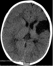
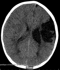
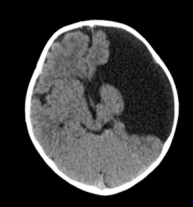
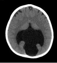
Presence of a left parietal cerebral cystic lesion with a density similar to CSF, well circumscribed, with polylobed contours, communicating with the subarachnoid space, exerting an attractive effect and dilation on the adjacent lateral ventricle. It is surrounded by cerebral parenchyma, which forms a hypodense ring compared to the rest of the parenchyma, possibly related to areas of gliosis (Figures 1 and 2).
There is no mass effect on the surrounding structures, which otherwise show no abnormalities in the supratentorial and infratentorial regions.
Contrast injection did not reveal any abnormal enhancement of the lesion or the rest of the parenchyma that could be related to meningoencephalitis.
Therefore, a diagnosis of porencephalic cyst was made.
4. Discussion
A porencephalic cyst or porencephaly is a rare condition. It is often congenital and discovered at birth.
[2]
S. Tambuzzi, G. Gentile, and R. Zoja, “Porencephalic cyst in adult,” Autops. Case Reports. 2022, vol. 12, pp. 10-13,
It can also be acquired in some cases and can vary in size, occupying both cerebral hemispheres, often respecting the territories supplied by the arteries of the circle of Willis.
[1]
A. T. Oommen, G. Sethy, N. T. Minz, J. Patra, and S. S. Panda, “Unusual presentation of porencephalic cyst in an adult,” J. Clin. Diagnostic Res. 2017, vol. 11, no. 2, pp. OD12-OD13,
Congenital brain lesions include two types of porencephaly:
1). Genetic porencephaly: resulting from poor development during early neuronal migration;
2). Encephalophaloplastic porencephaly, which is a late prenatal or perinatal vascular lesion due to an arterial ischaemic stroke or venous thrombosis.
[5]
A. Douzenis, E. N. Rizos, A. Papadopoulou, M. Papathanasiou, and L. Lykouras, “Porencephaly and psychosis: A case report and review of the literature,” BMC Psychiatry, 2010, vol. 10, pp. 2-5,
Some authors believe that the genetic theory supports the hypothesis of a mutant gene, COL4A1, which is thought to cause vascular wall damage leading to porencephaly in human families. In addition to this, there is often an environmental factor, which is usually trauma.
[6]
D. B. Gould et al., “Mutations in Col4a1 cause perinatal cerebral haemorrhage and porencephaly,” Science. May 2005, vol. 308, no. 5725, pp. 1167-1171,
The clinical presentation of porencephalic cysts varies widely, ranging from simple tonic-clonic seizures to motor deficits. The EEG may be normal or show focal rhythmic epileptiform discharges.
[7]
A. Qureshi, A. Jehangir, and E. P. York, “Porencephalic cyst: a rare cause of new-onset seizure in an adult,” J. Community Hosp. Intern. Med. Perspect., 2018, vol. 8, no. 2, pp. 92-93,
One study has claimed that the presence of a porencephalic cyst is not linked to recurrent episodes of epilepsy and that it is directly linked to hippocampal mesial sclerosis, which often coexists with congenital porencephalic cavities. They report the coexistence of amygdalo-hippocampal atrophy with unilateral or bilateral porencephalic cavities and that no cause-and-effect relationship has been established, but nevertheless recommend rigorous investigation for hippocampal atrophy whenever a porencephalic cyst is discovered.
[1]
A. T. Oommen, G. Sethy, N. T. Minz, J. Patra, and S. S. Panda, “Unusual presentation of porencephalic cyst in an adult,” J. Clin. Diagnostic Res. 2017, vol. 11, no. 2, pp. OD12-OD13,
Apart from the spastic crisis experienced by our patient, the symptoms depend on the area affected by the lesion and may manifest as psychiatric disorders, but the latter are more common in schizencephaly because it often affects the temporal lobe.
[5]
A. Douzenis, E. N. Rizos, A. Papadopoulou, M. Papathanasiou, and L. Lykouras, “Porencephaly and psychosis: A case report and review of the literature,” BMC Psychiatry, 2010, vol. 10, pp. 2-5,
A case of otorrhoea was described following a dehiscence of the tympanic membrane caused by this cystic mass, which was believed to be responsible for CSF leakage through the outer ear.
[8]
J.M Ryzenman, V.S. Rothholtz, R.J. Wiet “Porencephalic cyst: A review of the literature and management of a rare cause of cerebrospinal fluid otorrhea.” Otology and Neurotology, 2007, vol. 28, no. 3, pp. 381-386,
Another author reported the onset of diplopia and strabismus caused by a porencephalic cyst in the parieto-occipital region, the plausible mechanism being compression of the cerebral visual area.
[9]
A. H. Sarmast, H. I. Showkat, S. Farooq Mir, O. Masood, A. R. Kirmani, and A. R. Bhat, “Traumatic porencephaly with strabismus: A case report,” Iran. Red Crescent Med. J. 2012, vol. 14, no. 7, pp. 457-458.
[9]
.
Another author reported a case of a porencephalic cyst with intracerebral haemorrhage and deep vein thrombosis despite a negative thrombophilic assessment. The cause-and-effect relationship was not clearly established, but it was noted that the patient tested positive for a heterozygous mutation of the COL4A1 gene.
[10]
G. El Hasbani et al., “Intraparenchymal hemorrhage and cerebral venous thrombosis in an adult with congenital porencephalic cyst presenting for generalised tonic-clonic seizures,” Radiol. Case Reports, 2020, vol. 15, no. 1, pp. 95-99,
Given all this, we can see that porencephalic cysts can have a diverse clinical expression in the neurological sphere and that, in general, the treatment of porencephaly aims to relieve symptoms, as there is no treatment to induce brain growth in the missing sections; this may include anti-epileptic drugs, physiotherapy, or a shunt to remove excess cerebrospinal fluid.
The final diagnosis is made using imaging (CT or MRI). The CT scan shows a cystic lesion within a vascular territory, surrounded by cerebral parenchyma and communicating with either the lateral ventricle or the subarachnoid spaces.
[11]
P. Suthar Pokhraj, J. Patel Jigarj, M. Chetan, and A. Patel Narottama, “Congenital porencephaly in a new born child,” J. Clin. Diagnostic Res., 2014, vol. 8, no. 11, pp. RJ01-RJ02,
The advantage of MRI is that it provides the precision of the cerebral layer that delimits this cyst (white matter or grey matter), which distinguishes it from other entities that would constitute the range of differential diagnoses.
The differential diagnosis should be made with:
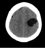
1). A neuroglial cyst: an intra- or extra-axial cystic formation in the central nervous system that does not communicate with the ventricle or subarachnoid spaces and is preferentially located in the frontal lobe. (Figure 3).
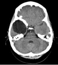
2). An arachnoid cyst: an extra-axial cyst that often exerts a mass effect on adjacent structures and is preferentially located in the temporal and occipital fossae. (Figure 4).
3). Schizencephaly: like porencephaly, it connects the subarachnoid space to the ventricle, which in this case is lined by the cerebral cortex or grey matter, unlike porencephaly, which is lined by white matter. (Figure 5).
4). Holoprosencephaly, which results from the failure of the hemispheres to separate, results in a single ventricle. (Figure 6)
[11]
P. Suthar Pokhraj, J. Patel Jigarj, M. Chetan, and A. Patel Narottama, “Congenital porencephaly in a new born child,” J. Clin. Diagnostic Res., 2014, vol. 8, no. 11, pp. RJ01-RJ02,
Figure 1. Well-defined left parietal porencephalic cavity communicating with the subarachnoid spaces.
Congenital porencephaly remains a little-known and rare condition in our environment, but it is responsible for a wide range of symptoms that can confuse clinicians and radiologists. Imaging (CT scan and MRI) remains essential for making a correct diagnosis. Prenatal morphological ultrasound should be rigorous and widely available in low-income countries in order to make an accurate early diagnosis of the malformation and guide the obstetrician-gynaecologist in the aetiological assessment and management of the newborn.
Obtained from the patient's mother, as the patient was a minor and was not able to consent on her own account.
Conflicts of Interest
The authors declare no conflicts of interest.
References
[1]
A. T. Oommen, G. Sethy, N. T. Minz, J. Patra, and S. S. Panda, “Unusual presentation of porencephalic cyst in an adult,” J. Clin. Diagnostic Res. 2017, vol. 11, no. 2, pp. OD12-OD13,
A. Nayak, S. Sharma, R. K. Vadher, S. Dixit, and R. S. Batra, “Congenital interparietal encephalocele: A case report,” J. Clin. Diagnostic Res. 2015, vol. 9, no. 4, pp. 9-10,
S. Tapadia, S. V. Phatak, H. G. K. B, and A. Pavanan, “Porencephalic Cyst in an Adult - A Rare Pathology,” J. Evol. Med. Dent. Sci. 2021, vol. 10, no. 12, pp. 918-919,
A. Douzenis, E. N. Rizos, A. Papadopoulou, M. Papathanasiou, and L. Lykouras, “Porencephaly and psychosis: A case report and review of the literature,” BMC Psychiatry, 2010, vol. 10, pp. 2-5,
A. Qureshi, A. Jehangir, and E. P. York, “Porencephalic cyst: a rare cause of new-onset seizure in an adult,” J. Community Hosp. Intern. Med. Perspect., 2018, vol. 8, no. 2, pp. 92-93,
J.M Ryzenman, V.S. Rothholtz, R.J. Wiet “Porencephalic cyst: A review of the literature and management of a rare cause of cerebrospinal fluid otorrhea.” Otology and Neurotology, 2007, vol. 28, no. 3, pp. 381-386,
A. H. Sarmast, H. I. Showkat, S. Farooq Mir, O. Masood, A. R. Kirmani, and A. R. Bhat, “Traumatic porencephaly with strabismus: A case report,” Iran. Red Crescent Med. J. 2012, vol. 14, no. 7, pp. 457-458.
[10]
G. El Hasbani et al., “Intraparenchymal hemorrhage and cerebral venous thrombosis in an adult with congenital porencephalic cyst presenting for generalised tonic-clonic seizures,” Radiol. Case Reports, 2020, vol. 15, no. 1, pp. 95-99,
P. Suthar Pokhraj, J. Patel Jigarj, M. Chetan, and A. Patel Narottama, “Congenital porencephaly in a new born child,” J. Clin. Diagnostic Res., 2014, vol. 8, no. 11, pp. RJ01-RJ02,
Marc, L., Dora, B., Sangwa-Milindi, Adelin, M., Gertrude, L. (2025). Congenital Porencephalic Cavity in a Newborn: Scanographic Aspect and Differential Diagnosis - A Case Report at Medpark Clinic. International Journal of Medical Imaging, 13(2), 27-30. https://doi.org/10.11648/j.ijmi.20251302.12
Marc, L.; Dora, B.; Sangwa-Milindi; Adelin, M.; Gertrude, L. Congenital Porencephalic Cavity in a Newborn: Scanographic Aspect and Differential Diagnosis - A Case Report at Medpark Clinic. Int. J. Med. Imaging2025, 13(2), 27-30. doi: 10.11648/j.ijmi.20251302.12
Marc L, Dora B, Sangwa-Milindi, Adelin M, Gertrude L. Congenital Porencephalic Cavity in a Newborn: Scanographic Aspect and Differential Diagnosis - A Case Report at Medpark Clinic. Int J Med Imaging. 2025;13(2):27-30. doi: 10.11648/j.ijmi.20251302.12
@article{10.11648/j.ijmi.20251302.12,
  author = {Lumbala-Mota Marc and Beyers-Mbayo Dora and Sangwa-Milindi and Muganza Adelin and Luyeye-Mvila Gertrude},
  title = {Congenital Porencephalic Cavity in a Newborn: Scanographic Aspect and Differential Diagnosis - A Case Report at Medpark Clinic
},
  journal = {International Journal of Medical Imaging},
  volume = {13},
  number = {2},
  pages = {27-30},
  doi = {10.11648/j.ijmi.20251302.12},
  url = {https://doi.org/10.11648/j.ijmi.20251302.12},
  eprint = {https://article.sciencepublishinggroup.com/pdf/10.11648.j.ijmi.20251302.12},
  abstract = {Aim: The objective of this study is to clarify the contribution of CT scan in the diagnosis of porencephalic cavities among the range of congenital cerebral cavities. Methods: Congenital porencephalic cavities are rare conditions characterised by the formation of a cyst in the cerebral parenchyma, which causes certain physical symptoms. We present the case of an 18-month-old infant who was referred to our clinic for sudden onset of right spastic hypotonia. Results: The CT scan revealed the presence of a left fronto-parietal cerebral cystic cavity communicating with the ipsilateral lateral ventricle, pointing to the characteristics of a congenital porencephalic cavity. The differential diagnosis included: - Neuroglial cyst;- Schizencephaly; - Arachnoid cyst;- Holoprosencephaly.Conclusion: Due to its rarity and atypical presentation, porencephaly poses a challenge for clinicians. The pathogenesis and treatment of this condition are poorly understood. Imaging is essential for establishing a diagnosis and determining the best treatment option. MRI is the modality of choice for characterising cerebral cystic lesions. In countries with limited resources, CT scanning is a valuable tool for characterising lesions while ruling out other diagnoses.},
 year = {2025}
}
TY  - JOUR
T1  - Congenital Porencephalic Cavity in a Newborn: Scanographic Aspect and Differential Diagnosis - A Case Report at Medpark Clinic

AU  - Lumbala-Mota Marc
AU  - Beyers-Mbayo Dora
AU  - Sangwa-Milindi
AU  - Muganza Adelin
AU  - Luyeye-Mvila Gertrude
Y1  - 2025/07/31
PY  - 2025
N1  - https://doi.org/10.11648/j.ijmi.20251302.12
DO  - 10.11648/j.ijmi.20251302.12
T2  - International Journal of Medical Imaging
JF  - International Journal of Medical Imaging
JO  - International Journal of Medical Imaging
SP  - 27
EP  - 30
PB  - Science Publishing Group
SN  - 2330-832X
UR  - https://doi.org/10.11648/j.ijmi.20251302.12
AB  - Aim: The objective of this study is to clarify the contribution of CT scan in the diagnosis of porencephalic cavities among the range of congenital cerebral cavities. Methods: Congenital porencephalic cavities are rare conditions characterised by the formation of a cyst in the cerebral parenchyma, which causes certain physical symptoms. We present the case of an 18-month-old infant who was referred to our clinic for sudden onset of right spastic hypotonia. Results: The CT scan revealed the presence of a left fronto-parietal cerebral cystic cavity communicating with the ipsilateral lateral ventricle, pointing to the characteristics of a congenital porencephalic cavity. The differential diagnosis included: - Neuroglial cyst;- Schizencephaly; - Arachnoid cyst;- Holoprosencephaly.Conclusion: Due to its rarity and atypical presentation, porencephaly poses a challenge for clinicians. The pathogenesis and treatment of this condition are poorly understood. Imaging is essential for establishing a diagnosis and determining the best treatment option. MRI is the modality of choice for characterising cerebral cystic lesions. In countries with limited resources, CT scanning is a valuable tool for characterising lesions while ruling out other diagnoses.
VL  - 13
IS  - 2
ER  -