Introduction: Acetabular fractures are relatively rare injuries. The purpose of this study was to evaluate the functional and radiographical outcomes of surgical management of Acetabular fractures in medium term. Materials and method: This was a retrospective and descriptive study conducted on the records of patients operated on for acetabular fractures classified according to Letournel and Judet criteria between January 1, 2021, and December 31, 2024. Clinical and imaging examinations were used to establish the diagnosis and indicate surgery. Anteroposterior pelvic radiographs were used to assess joint congruity according to Duquesnoy and Senegas criteria, reduction according to Matta criteria, and bone healing. Functional assessment was based on the Postel-Merle d’Aubigne criteria. Results: The series included 15 patients (15 fractures) (13 men and 2 women). The mean age was 37.8 (23 and 55) years. The fracture was due to road traffic accident (n=13) and fall from height (n=2). The fracture involved the posterior wall (n=3), posterior column (n=3), anterior column (n=2), transverse (n=1), T-shaped (n=1), associated transverse and posterior wall (n=2), posterior column and posterior wall (n=2) and bicolumn (n=1). The mean time to surgery was 13.1 (8 and 20) days. The mean hospital stay was 24 (18 and 40) days. The mean follow-up was 2.8 years (8 months and 4 years). Head/roof congruence was perfect (n=12), good (n=2), and fair (n=1). Head/acetabulum congruence was perfect in all cases. Reduction was anatomical (n=12), satisfactory (n=2), and unsatisfactory (n=2). The Postel-Merle d’Aubigne functional score was excellent (n=8), good (n=5), and fair (n=2). Conclusion: Acetabular fractures are relatively rare injuries. They occur following high-energy trauma. Surgical management within a short timeframe, using an appropriate approach, often results in anatomic reduction and a congruent hip. The functional outcome is satisfactory in the medium term.
| Published in | American Journal of Orthopaedics and Traumatology (Volume 1, Issue 1) |
| DOI | 10.11648/j.ajot.20260101.12 |
| Page(s) | 9-13 |
| Creative Commons |
This is an Open Access article, distributed under the terms of the Creative Commons Attribution 4.0 International License (http://creativecommons.org/licenses/by/4.0/), which permits unrestricted use, distribution and reproduction in any medium or format, provided the original work is properly cited. |
| Copyright |
Copyright © The Author(s), 2026. Published by Science Publishing Group |
Acetabular Fracture, Joint Congruity, Surgical Treatment
Head/roof congruence | Preoperative | Postoperative |
|---|---|---|
Bad | 5 | 0 |
Fair | 7 | 1 |
Good | 3 | 2 |
Perfect | 0 | 12 |
congruence of head/acetabulum | Preoperative | Postoperative |
|---|---|---|
Bad | 5 | 0 |
Fair | 3 | 0 |
God | 4 | 0 |
Perfect | 3 | 15 |
SOFCOT | French Society of Orthopaedic and Trauma Surgery |
SPSS | Statistical Package for Social Sciences |
PMA | Postel-merle d’Aubigne |
| [1] | Rahimi H, Gharahdaghi M, Parsa A, Assadian M. Surgical Management of Acetabular Fractures: A Case Series. Trauma Mon. 2013; 18(1): 28–31. |
| [2] | Yang Y, Yi M, Zou C, Yan ZK, Yan XA, Fang Y. Mapping of 238 quadrilateral plate fractures with three-dimensional computed tomography. Injury. 2018; 49(7): 1307–12. |
| [3] | Labsaili N, Rahmi M, Bekkali Y, and al. management of acetabular fractures (6 year setback). Rev Maroc Chir Orthop Traumato 2009; 40: 35-39. |
| [4] | Mauffrey C, Hao J, Cuellar DO, and al. The epidemiology and injury patterns of acetabular fractures: Are the USA and China comparable? Clin Orthop Relat Res 2014; 472: 3332‐7. |
| [5] | Guerado E, Cano JR, Cruz E. Fractures of the acetabulum in elderly patients: An update. Injury 2012; 43(2): 33‐41. |
| [6] | Baba HF, Oudrhiri D, Benchekroun S, and al. Surgical management of acetabular fractures: experience of the orthpedic trauma department B4, university hospital Hassan II. IOSR-JDMS. 2020; 19(11): 42-45. |
| [7] | SOFCOT collective. Surgical management in displaced acetabular fractures. Teaching conferences. Paris: Elsevier; 2015. p. 203-215. |
| [8] | Ben Salah M, Karray S, Frikha I, and al. Acetabular fractures in the elderly. Tunis Med. 2020; 98(3): 189-195. |
| [9] | Fakru N, Faisham W, Hadizie D, Yahaya S. Functional Outcome of Surgical Stabilisation of Acetabular Fractures. Malays Orthop J. 2021; 15(2): 129–35. |
| [10] | Ramesh KS, Sujit KT, Sameer A, Tarun G, Dharm SM, Santosh M. A safe technique of anterior column lag screw fixation in acetabular fractures.’ International orthopaedics 36.11 (2012): 2333-2340. |
| [11] | Etemadifar M, Nemati A, Chinigarzade M. Operative management of acetabular fracture: A 10-year experience in Isfahan, Iran. Adv Biomed Res. 2016; 5: 169. |
| [12] | Judet R, Letournel E. General principales of management of acetabular fractures. Fractures of acetabulum. Ed 1993 ; Springer, Berlin Heidelberg, New York. |
| [13] | Matta JM, Mehne DK, Roffi R. Fractures of the acetabulum. Early results of a prospective study. Clin Orthop. 1986; 205: 241–50. |
| [14] | Matta JM, Merritt PO. Displaced acetabular fractures. Clin Orthop. 1988; 230: 83–97. |
| [15] | Routt ML, Swiontkowski MF. Operative treatment of complex acetabular fractures. Combined anterior and posterior exposures during the same procedure. J Bone Joint Surg Am. 1990; 72(6): 897–904. |
| [16] | Judet R, Judet J, Letournel E. Fractures of the acetabulum: Classification and surgical approaches foropen reduction. Preliminary report. J Bone Joint Surg Am. 1964; 46: 1615–46. |
| [17] | Duquennoy A, Tillie B, Delcourt JP. Acetabular fractures : Joint congruence and therapeutic indication. Acta Orthop Belg 1984 ; 50(3) : 343-55. |
| [18] | Merle d’Aubigne R, Postel M. Functional results of hip arthroplasty with acrylic prosthesis. J Bone Joint Surg Am. 1954; 36: 451-75. |
| [19] | Rkiba Z, Rajaallah A, Sidi El, Rafai M, Rahmi M, Garch A. Surgical treatment of acetabular fratures: Predictive factors of medium and long term outcome. SMACOT. 2020; 88: 17-24. |
| [20] | Rahmi M, Arssi M, Doumane B, Cohen D, Fnini S, The value of computed tomography in acetabular fractures: A study of 30 cases. Maghreb Med 2001; 21(359).335-37. |
| [21] | Kelly J, Ladurner A, Rickman M. Surgical management of acetabular fractures e a contemporary literature review. Injury. 2020; 1383(20): 30525-30528. |
| [22] | Letournel E. Acetabulum fractures: classification and management. Clin Orthop Relat Res. 1980; 151: 81-106. |
| [23] | Judet R, Louternel E. Acetabular fractures. Masson. Paris. 1974. |
| [24] | Mahdane H, Elghazi A, Shimi M, Elibrahimi A, Elmrini A. Surgical treatment of acetabular fractures: A report of 22 cases. Pan Afr Med J. 2014; 17: 123.1-6. |
| [25] | Olson SA, Guilak F. From Articular Fracture to Posttraumatic Arthritis: A Black Box That Needs to Be Opened. J Orthop Trauma. 2006; 20(10): 661–2. |
| [26] | Duquennoy A, Senegas J, Augereau B, et al. [Fractures of the acetabulum. Results 5 years later. Round table]. Rev Chir Orthop Reparatrice Appar Mot. 1982; 68(2): 45–82. |
| [27] |
Ziran N, Soles GLS, Matta JM (2019) Outcomes after surgical treatment of acetabular fractures: a review. Patient Saf, Surg, p 13.
https: //doi.org/10.1186/s13037-019-0196-2 |
| [28] | Murphy D, Kaliszer M, Rice J, McElwain JP. Outcome after acetabular fracture. Prognostic factors and their inter-relationships. Injury 2003; 34: 512—7. |
| [29] | Vipulendran K, Kelly J, Rickman M, Chesser T. Current concepts: managing acetabular fractures in the elderly population. Eur J Orthop Surg Traumatol. 2021; 31(5): 807–16. |
| [30] |
Briffa N, Pearce R, Hill AM, Bircher M. Outcomes of acetabular fracture fixation with ten years’ follow-up. J Bone Joint Surg Br. 2011; 93(2): 229–36.
https: //doi.org/10.1302/0301-620X.93B2.24056 |
| [31] | Goulet JA, Bray TJ. Complex acetabular fractures. Clin Orthop. 1989; 240: 9–20. |
APA Style
Korka, D. M., Amadou, B. P., Sekou, C. M., Moustapha, D. M., Diouf, N. C. (2026). Surgical Management of Acetabular Fractures in Adultes: About 15 Cases. American Journal of Orthopaedics and Traumatology, 1(1), 9-13. https://doi.org/10.11648/j.ajot.20260101.12
ACS Style
Korka, D. M.; Amadou, B. P.; Sekou, C. M.; Moustapha, D. M.; Diouf, N. C. Surgical Management of Acetabular Fractures in Adultes: About 15 Cases. Am. J. Orthop. Traumatol. 2026, 1(1), 9-13. doi: 10.11648/j.ajot.20260101.12
@article{10.11648/j.ajot.20260101.12,
  author = {Diallo Mamadou Korka and Ba Papa Amadou and Conde Mamady Sekou and Diallo Mamadou Moustapha and Niang Coumba Diouf},
  title = {Surgical Management of Acetabular Fractures in Adultes: About 15 Cases},
  journal = {American Journal of Orthopaedics and Traumatology},
  volume = {1},
  number = {1},
  pages = {9-13},
  doi = {10.11648/j.ajot.20260101.12},
  url = {https://doi.org/10.11648/j.ajot.20260101.12},
  eprint = {https://article.sciencepublishinggroup.com/pdf/10.11648.j.ajot.20260101.12},
  abstract = {Introduction: Acetabular fractures are relatively rare injuries. The purpose of this study was to evaluate the functional and radiographical outcomes of surgical management of Acetabular fractures in medium term. Materials and method: This was a retrospective and descriptive study conducted on the records of patients operated on for acetabular fractures classified according to Letournel and Judet criteria between January 1, 2021, and December 31, 2024. Clinical and imaging examinations were used to establish the diagnosis and indicate surgery. Anteroposterior pelvic radiographs were used to assess joint congruity according to Duquesnoy and Senegas criteria, reduction according to Matta criteria, and bone healing. Functional assessment was based on the Postel-Merle d’Aubigne criteria. Results: The series included 15 patients (15 fractures) (13 men and 2 women). The mean age was 37.8 (23 and 55) years. The fracture was due to road traffic accident (n=13) and fall from height (n=2). The fracture involved the posterior wall (n=3), posterior column (n=3), anterior column (n=2), transverse (n=1), T-shaped (n=1), associated transverse and posterior wall (n=2), posterior column and posterior wall (n=2) and bicolumn (n=1). The mean time to surgery was 13.1 (8 and 20) days. The mean hospital stay was 24 (18 and 40) days. The mean follow-up was 2.8 years (8 months and 4 years). Head/roof congruence was perfect (n=12), good (n=2), and fair (n=1). Head/acetabulum congruence was perfect in all cases. Reduction was anatomical (n=12), satisfactory (n=2), and unsatisfactory (n=2). The Postel-Merle d’Aubigne functional score was excellent (n=8), good (n=5), and fair (n=2). Conclusion: Acetabular fractures are relatively rare injuries. They occur following high-energy trauma. Surgical management within a short timeframe, using an appropriate approach, often results in anatomic reduction and a congruent hip. The functional outcome is satisfactory in the medium term.},
 year = {2026}
}
TY  - JOUR
T1  - Surgical Management of Acetabular Fractures in Adultes: About 15 Cases
AU  - Diallo Mamadou Korka
AU  - Ba Papa Amadou
AU  - Conde Mamady Sekou
AU  - Diallo Mamadou Moustapha
AU  - Niang Coumba Diouf
Y1  - 2026/01/29
PY  - 2026
N1  - https://doi.org/10.11648/j.ajot.20260101.12
DO  - 10.11648/j.ajot.20260101.12
T2  - American Journal of Orthopaedics and Traumatology
JF  - American Journal of Orthopaedics and Traumatology
JO  - American Journal of Orthopaedics and Traumatology
SP  - 9
EP  - 13
PB  - Science Publishing Group
UR  - https://doi.org/10.11648/j.ajot.20260101.12
AB  - Introduction: Acetabular fractures are relatively rare injuries. The purpose of this study was to evaluate the functional and radiographical outcomes of surgical management of Acetabular fractures in medium term. Materials and method: This was a retrospective and descriptive study conducted on the records of patients operated on for acetabular fractures classified according to Letournel and Judet criteria between January 1, 2021, and December 31, 2024. Clinical and imaging examinations were used to establish the diagnosis and indicate surgery. Anteroposterior pelvic radiographs were used to assess joint congruity according to Duquesnoy and Senegas criteria, reduction according to Matta criteria, and bone healing. Functional assessment was based on the Postel-Merle d’Aubigne criteria. Results: The series included 15 patients (15 fractures) (13 men and 2 women). The mean age was 37.8 (23 and 55) years. The fracture was due to road traffic accident (n=13) and fall from height (n=2). The fracture involved the posterior wall (n=3), posterior column (n=3), anterior column (n=2), transverse (n=1), T-shaped (n=1), associated transverse and posterior wall (n=2), posterior column and posterior wall (n=2) and bicolumn (n=1). The mean time to surgery was 13.1 (8 and 20) days. The mean hospital stay was 24 (18 and 40) days. The mean follow-up was 2.8 years (8 months and 4 years). Head/roof congruence was perfect (n=12), good (n=2), and fair (n=1). Head/acetabulum congruence was perfect in all cases. Reduction was anatomical (n=12), satisfactory (n=2), and unsatisfactory (n=2). The Postel-Merle d’Aubigne functional score was excellent (n=8), good (n=5), and fair (n=2). Conclusion: Acetabular fractures are relatively rare injuries. They occur following high-energy trauma. Surgical management within a short timeframe, using an appropriate approach, often results in anatomic reduction and a congruent hip. The functional outcome is satisfactory in the medium term.
VL  - 1
IS  - 1
ER  -
Traumatology and Orthopedics Department, Cheikh Anta Diop University, Dakar, Senegal
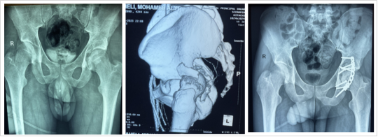
Figure 1. Radiographical Aspect of Acetabular Posterior Wall Fracture Managed with a Posterior Plate.
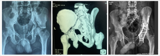
Figure 2. Radiographical Aspect of Acetabular Bicolumnar Fracture Managed with Anterior and Posterior Plates.
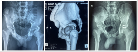
Figure 3. Radiographical Aspect of Acetabular Posterior Wall Fracture Managed by Screw Fixation.
Information